ORGAN OF HEARING Ear
Organ of Hearing or Organ of equilibrium, Ear Structure and Function
Dr Pramila Singh
2/24/20245 min read

HSBTE DMLT IInd Semester Organ of Hearing or Organ of Equilibrium: Ear Structure and Function
ORGAN OF HEARING or Organ of equilibrium
Hearing is the detection of sound mechanical waves (Vibration). It converts sound vibration into the sensory impulse to be understood by the temporal lobe in the brain.
The organ of hearing and the organ of equilibrium is the ear. Ear responds to sound and similar vibrations thus it is also called Phonoreceptor.
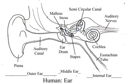
Structure of the Human ear: The human ear consists of three parts:
a. External ear
b. Middle ear
c. Inner ear.
A. EXTERNAL EAR:
The external ear consists of three parts: Pinna, External Auditory Canal, and Tympanum
1. Pinna (Auricle or ear lobe): Pinna is made of elastic cartilage covered by skin. Pinna has a soft prominent outer ridge known as helix and a soft vascular lower part made of fibrous and adipose tissues called lobule. The depression part of the pinna is called concha. Pinna collects sound waves and directs them into the external auditory canal.
2. External auditory canal (external auditory meatus): Concha of the pinna leads to the external auditory canal. The external auditory canal is ‘S-shaped’, irregular tubular canal ends at the tympanum.
Its external part is cartilaginous and the internal part is bony. The outer part of the canal has hairs on the skin. Wax glands (ceruminous glands) are present on the inner portion of the canal. The Wax gland secretes ear wax (cerumen). Hair and wax protect the tympanum from dust, microbes, and changes in temperature, humidity, and water during bathing/swimming. etc.
3. Tympanum (Eardrum): Tympanum is between the external and middle ear. It is also called the eardrum or tympanic membrane.
· It is a translucent oval membrane that covers the tympanic cavity. The outer surface is slightly concave and the edge is slightly thick.
· It consists of three layers. The outer layer, middle layer, and inner layer.
· The inner layer is rich in nerve fibers and blood vessels. These nerves make it sensitive to pain.
· The tympanic membrane has a depressed part in the center on the inner surface (Toward the middle ear). It is called the umbo.

. MIDDLE EAR:
It consists of the tympanic cavity and ear ossicles
1. Tympanic Cavity: It is present in the temporal bone, filled with air. The tympanic cavity is connected with the nasopharynx through the auditory tube (Eustachian tube
· Eustachian tube air equalizes air pressure on both sides of the tympanic membrane (inner side in the tympanic cavity and outside in the external ear).
· . Eustachian tube opening remains closed during normal conditions. It opens to equalize middle ear air pressure and external ear air pressure (atmospheric pressure).
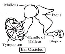
2. Ear Ossicles: The tympanic cavity has three flexible small bones called ear ossicles (auditory ossicles). These are Malleus, Incus and Stapes.
· Malleus is hammer-shaped one end attached to the inner side of the tympanic membrane and the other end with incus.
· . The incus is an anvil-shaped middle bone connected with a malleus at one end and with stapes at the other end.
· Stapes is a stirrup-shaped inner bone attached to the incus at one end and with an oval window of the inner ear at another end. Stapes are the smallest bones of the human body. The malleus is the largest bone among all three ear ossicles.
The middle ear opens into the internal ear through two apertures. Both apertures are covered with a membrane. These apertures are fenestra ovalis (oval window) and fenestra rotunda (Round window).
Middle ear function: Sound waves vibrate the tympanic membrane causing vibration in the middle ear. Middle ear vibration transmits sound waves from the external ear to the internal ear through ear ossicles.


A. INNER EAR:
The temporal bone forms the internal ear. The Internal ear is a cavity lined with a membrane called a Labyrinth. Labyrinth floats on the fluid present in labyrinth cavities. Fluid in the labyrinth is called perilymph. Perilymph composition is the same as cerebrospinal fluid (CSF). The labyrinth consists of the vestibule, semicircular canals, and cochlea.
Vestibule: Vestibule consists of the following
1. Utriculus and Sacculus: The Vestibule has two sacs upper larger utriculus and a lower smaller sacculus. Both are connected with the utricula-saccular duct.
2. Ductus: The posterior end of the utricular-saccular duct has an elongated tube called ductus endolymphaticus. The lower end of the saccule has a short tube called ductus reuniens. Ductus reuniens is connected with the cochlear duct.
3. Maculae: Both saccule and utricle have a sensory spot called maculae. The macula consists of sensory cells and supporting cells.
4. Sensory cells: Sensory cells have various non-motile hairs (microvilli) and one cilium. Maculae help to maintain the static balance of the body.
Semicircular canal: It consists of the following
1. Labyrinth: The labyrinth has three semicircular canals: anterior semicircular canal, Posterior semicircular canal, and lateral semicircular canal.
2. Ampulla: Each canal is enlarged at one end called the ampulla.
3. Cristae (Singular: Crista): Each ampulla has a sensory nerve cell area called crista. Each crista has sensory nerve cells and supporting cells. Sensory cells have various nonmotile hairs and a long cilium. Cristae are associated with the balance of the body.
The semicircular canal is associated with the movement of the head. It forwards signals to the brain. The brain senses the rate of head movement and the direction of head movement.
Cochlea: It is a spiral twisted tube. It consists of the following
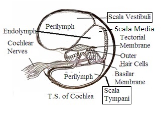
1. Scala: Internally cochlea has three chambers filled with fluid. These chambers are the upper scala vestibule, lower scala tympani, and middle scala media (cochlear duct).
2. Scala fluids: Fluid in the scala vestibule and scala tympani is called perilymph and scala media (cochlear duct) is filled with endolymph.
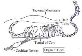
3. Organ of Corti: Scala media floor (basilar membrane) has sensory cells called organ of Corti. The organ of Corti is the sound receptor organ.

THE FUNCTION OF THE EAR:
Hearing and 2. equilibrium
Mechanism of Hearing:
1. Pinna collects sound waves and passes them to the tympanic membrane. Sound waves vibrate the tympanic membrane.
2. Vibrations from the tympanic membrane are transmitted to ear ossicles (malleus, incus, and stapes bone). Ear ossicles amplify sound waves vibration by about 20 times. It further vibrates perilymph.
3. Perilymph vibrations are transmitted to the vestibule and scala. This vibration stimulates the vibration of the endolymph.
4. Endolymph vibration stimulates the organ of Corti sensory hairs. Organ of Corti sensory hairs (sensory photoreceptors) generate sensory impulses.
5. These sensory impulses are transmitted to the brain temporal lobe of both cerebral hemispheres. Temporal lobes recognize the hearing sensation.
Mechanism of Equilibrium: Cristae and maculae maintain the equilibrium of the body.
Cristae: Cristae maintain the dynamic equilibrium of the body
1. Hair cells of cristae sense body movement and send signals to the brain.
2. The medulla oblongata of the brain detects the change in position. The brain transmits the motor impulse to body muscles to correct the position.
3. Long travel dizziness, spinning dizziness, etc are due to excessive sensitization and continuous disturbance in the endolymph.
Maculae: Maculae maintain the static equilibrium of the body
1. Change in head position generates sensory impulses that are transmitted to the cerebellum in the brain.
2. The cerebellum transmits the sensory impulse to maintain a static position.
3. The Semicircular canal, utricle, and saccule are considered as a structure of equilibrium or a structure of balancing. These are part of the labyrinth.
Ear Diseases
Otitis media: Inflammation in the middle ear is called otitis.
Balance disturbance: Operation in-ear, traveling (traveling sickness or motion sickness) may develop dizziness. Injury of the head may permanently affect body gait and body balance.
Dr. Pramila Singh.
