Nervous System
HSBTE DMLT IInd Semester Nervous system 1.1 Central nervous system (brain and spinal cord) 1.2 Peripheral nervous system (cranial and spinal nerves) 1.3 The sense organs (eye, ear, tongue, and nose); structure and functions
Dr Prmila Singh
2/18/20247 min read

HSBTE DMLT IInd Semester Nervous system: Central nervous system (brain and spinal cord), Peripheral nervous system (cranial and spinal nerves), The sense organs (Eye, Ear, Tongue, and Nose); structure and functions
UNIT NERVOUS SYSTEM
Introduction
The nervous system is also called a neural system that coordinates various functions of the body parts. The neural system has mainly three basic functions:
1. To receive impulses from organs,
2. To process and analyze impulses,
3. To send impulses to control the organ's activities.
There are two types of Nervous System (neural systems)
A. Central Nervous System (CNS)
B. Peripheral Nervous System (PNS)
· The Central Nervous System consists of the brain inside the cranium (skull) and the spinal cord inside the vertebral column.
· The peripheral nervous system consists of nerves and ganglia. Nerves arise from the brain and spinal cord.
· Nerves that arise from the brain are called the cranial nerves and nerves that arise from the spinal cord are called spinal nerves.
· Spinal nerves spread laterally from the spinal cord throughout the body. Cranial nerves run from the brain to the spinal cord.
The Peripheral Nervous System (PNS) has two types of nerve fibers depending on their functions. These are
·1. Afferent nerve fibers or sensory nerve fibers: They carry sensory impulses from tissues or organs to the central nervous system (CNS).
· 2. Efferent nerve fibers or motor nerve fibers: They carry motor nerve impulses from CNS to effecter organs.
There are two types of peripheral nervous system (PNS)
1. Somatic nervous system: The somatic nervous system transmits the impulse from the brain to skeletal muscles, joints, bones, ligaments, skin, eyes, ears, etc, and from these organs to the brain. The movement of these parts is under the control of human will that is the voluntary movement of organs and body parts.
2. Visceral nervous system: The visceral nervous system transmits impulses to the brain from visceral organs muscles like cardiac muscles and smooth muscles and from them to the brain. These movements are involuntary. It is also called the autonomic nervous system (ANS).
The autonomic nervous system is again divided into two parts
A. Sympathetic nervous system
B. Parasympathetic nervous system
Central Nervous System
The central nervous system consists of the Brain (Encephalon) and Spinal Cord.
Brain (Encephalon)
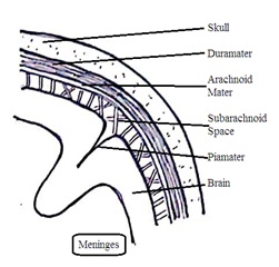
The brain is the uppermost of the central nervous system present in the cranial cavity (cranium) of the skull. The average brain weight is 1.2 kg to 1.4 kg. It is covered by maninges made of three layers Piamatter, Duramatter, and Arachnoid space.
1. Piamatter: It is the innermost, thin, vascular, tough pigmented layer covered. The area of pia matter and brain fusion is called the choroid plexus. The Choroid plexus is found on the roof of the brain.
2. Arachnoid matter: It is the thin, middle, webby, porous, nonvascular layer. The space between arachnoid matter and pia matter is called subarachnoid space. It is filled with connective tissues and cerebrospinal fluid (CSF). CSF protects the brain from shock. It also provides nutrition, respiratory gases, etc to the brain and removes toxic or waste materials from the brain.
3. Duramatter: It is the outermost nonvascular, thick, hard, double-layered fibrous tissue of the brain. It remains in close contact with the inner side of the skull.. The space between dura matter and arachnoid matter is called subdural space. Subdural space is filled with fluid but it is not cerebrospinal fluid (CSF).


The brain is made of two types of cells. These are gray matter and white matter. Gray matter is the external part of the brain that consists of the neuron cell body, dendrites, and non-medullated nerve fibers. It is grey.
White matter is an internal part of the brain that consists of medullated nerve fibers. Thus in white matter nerve impulses transmit more rapidly than in gray matter.
The human brain has three parts the Fore-brain, Mid-brain, and Hind-brain
A. Fore-brain (Prosencephalon): It is the front part of the brain and consists of the olfactory lobe, cerebrum, and diencephalon.
· 1. Olfactory lobe (Rhinocephalon): There are two clubbed-shaped olfactory lobes in the brain.Olfactory nerves arise from olfactory lobes. The olfactory lobe is the sense organ of smell.
· 2. Cerebrum: It is the largest part of the brain made of two cerebral hemispheres. These are the left and right hemispheres. On the inferior side, they are connected by the corpus callosum. The left side of the cerebrum controls the right side of the body and the right side of the cerebrum controls the right side of the body.
3. Cerebral Cortex: The cerebral cortex is the outer portion of the cerebrum in the gray matter area. The cerebral cortex has three regions.
· Sensory region: Nerve fibers arise from the sensory region and are responsible for sensation, hearing, and vision.
· Motor region: Nerves from the motor region are for locomotion and movement
· Association region: Stores impulse input and initiates response.
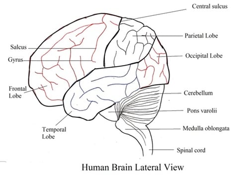
.Each cerebrum has four lobes.
· Frontal lobe: They control voluntary muscle contraction. Motor speech area is present in the frontal lobe
· Parietal lobe: It receives pain, temperature, pressure, and touch sensory sensation.
· Temporal lobe: The temporal lobe has a sensory speech area. It extends from the temporal lobe to the posterior parietal lobe. The speech area is present in the lower part of the parietal lobe and the auditory area is present in the temporal area below the lateral sulcus (lateral fissure).
· Occipital lobe: The visual area and visual sensation area are present in the occipital lobe. occipital lobe.
4. Diencephalon: It consists of the epithalamus, thalamus, and hypothalamus.
· Epithalamus is connected with the endocrine gland pineal body.
· Thalamus is present just above mid brain. The hypothalamus is attached to the pituitary gland through the infundibulum.
· The hypothalamus is highly vascular. and maintains homeostasis.
Ventricles of Brain: The brain has four hollow liquid-filled irregular spaces called ventricles. Two lateral ventricles, one-third ventricle, and one-fourth ventricle. CSF moves in one direction to subarachnoid space.
B. Midbrain (Mecencephalon): Midbrain is a group of nerve cells present in the white matter of the brain.
Mid brain consists of two structures corpora quadrigemnia and crura cerebri.
1. Corpora Quadrigemnia: The upper surface of the midbrain has two rounded swellings on each side. These four rounded protrusions (knob or swelling) are called corpora Quadrigemnia. One pair of quadrigemnia is called corpora bigemnia. Out of four corpora quadrigeminal two are superior and two are inferior in position
· Superior caliculi are related to vision to receive impulses from eyes and head muscles. This helps to regulate eye movement and head movement to locate objects.
· Inferior colliculi are related to hearing to receive impulses from ear and head muscles. This helps to regulate the ear and to detect sound objects.
2. Crura Cerebri (cerebral peduncle): The lower surface of the midbrain has two bundles of fibers called cerebral peduncles. Impulses from the cerebrum, cerebellum, pons, and medulla are relayed through crura cerebri.
C. Hindbrain (Metencephalon): It consists of the cerebellum, pons verolii, and medulla oblongata.
1. Cerebellum (Epencephalon): Cerebellum means little cerebrum. It has three parts. These are two lateral cerebellum hemispheres and one middle vermis. The cerebellum is present in between the cerebrum and medulla oblongata. It has three cerebellar peduncles. These are Superior, Middle, and Inferior.
· Impulses travel from the superior cerebellum peduncle to the midbrain, middle cerebellum peduncle to pons, and inferior cerebellum peduncle to the medulla oblongata and spinal cord.
· Cerebellum regulates various muscular activities like running, typing, talking, cycling, etc. It also maintains the equilibrium of body and muscle tone.
2. Pons Verolii: It is present in between the medulla oblongata and mid-brain, in front of the cerebellum. It connects both hemispheres of the cerebellum, cerebrum, medulla oblongata, and spinal cord.
3. Medulla oblongata: It is present in between pons verolii and the spinal cord. It is the lowermost important part of the brain that encloses the fourth ventricle. Some part of the medulla oblongata has no nervous tissue called the posterior choroid plexus. It helps to synthesize cerebrospinal fluid (CSF).
· It receives impulses from the spinal cord and passes these impulses to the cerebellum and thalamus.
· It regulates heart rate, blood pressure, coughing, sneezing, swallowing, salivation, breathing, and various involuntary activities.
SPINAL CORD
It is the posterior part (lower part) of the central nervous system present inside the vertebral column. It is protected by meninges. Meninges consist of Piamater, arachnoid mater, and Dura mater.
1. The Piamater is a thin, innermost, highly vascular pigmented layer with squamous epithelium.
2. The arachnoid mater is the thin, middle, non-vascular layer with squamous epithelium. The space between the arachnoid mater and the pia mater is called sub-arachnoid space. Subarachnoid space is filled with cerebrospinal fluid.
3. The Dura mater is a thick outermost strong layer made of fibrous connective tissues. The space between the dura mater and vertebral column is called epidural space. Epidural space is filled with connective tissues, adipose tissues, and blood veins. This space acts as a cushion to absorb shock.
The spinal cord does not secrete CSF. CSF enters the spinal cord from the brain. The spinal cord extends from the brain medulla oblongata to the second lumbar vertebrae. It is curved and shaped with varying diameters.
· One curvature is at shoulder level (Cervical region), and the other curvature is at hip level (lumbar region). These curvatures have a maximum number of nerve cells and they are enlarged areas.
· The cervical region spinal cord supplies nerves to arms and the lumbar region spinal cord supplies nerves to legs.
· Cervical region spinal cord enlargement is present between the fourth cervical vertebrae and thoracic vertebrae region. Lumbar region spinal cord enlargement is in the abdominal region.
· The spinal cord end has tapered. This tapered end is called Conus medullaris.
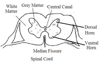
· Internally posterior median sulcus and anterior median sulcus divide the spinal cord into two halves (Right and left halves).
· The external area of the spinal cord is made of white matter. White matter is a bundle of myelinated nerve fibers called fascicule.
· Bundles of axons arise from the fascicule to the brain. This is called the ascending tract. This carries sensory impulses from the spinal cord to the brain. Similarly, the descending tract arises from the brain to the spinal cord. This carries impulses from the brain to the spinal cord.
· White matter surrounds grey matter present in the internal area of the spinal cord. Grey matter is a bundle of non-myelinated nerve fibers. Grey matter forms a butterfly-shaped area in the spinal cord.
· Six horns arise from grey matter. These are two lateral horns, two anterior horns (ventral horn), and two posterior horns (dorsal horn). The spinal cord has a central canal containing cerebrospinal fluid.
Functions: The spinal cord is the center of the spinal reflex. Sensory impulses from all parts of the body to the brain pass through the spinal cord. Similarly, all motor impulses from the brain to body parts pass through the spinal cord.
Dr Pramila Singh.


Peripheral Nervous system (Cranial nerves and Spinal nerves)
Nerves originate from the central nervous system to connect receptor organs to form the peripheral neural system. There are two types of nerves in the peripheral neural system. These are Cranial nerves and spinal nerves.
Cranial Nerve (Cerebral Nerve)
Nerves arise from the brain and pass through various openings of the cranial bones are called cranial nerves. The human peripheral neural system has 12 pairs of cranial nerves.
1. Olfactory Nerve (Cranial Nerve I): Nerve fibers arise from olfactory epithelium in the nasal chamber. It passes through the olfactory bulb and runs through the olfactory tract to terminate in the temporal lobe of the cerebrum. It is the sensory nerve of smell.
2. Optic Nerve (Cranial Nerve-II): Nerve fibres arise from in retina of the eyes and combine to form optic nerve. Optic nerve fibers enter the occipital lobe of the brain. The optic nerve is the sensory nerve of sight.
3. Oculomotor Nerve (Cranial Nerve III): Nerve fibres arise from mid brain floor and terminate in ocular muscles. It is motor nerves to control eyeball rotation, pupil size, and accommodation of eyes.
4. Pathetic or Trochlear Nerve (Cranial Nerve IV): It arises from the mid-brain floor and terminates in eye eyeball oblique muscles. It is a motor nerve to controls eyeball movement. It is the thinnest and smallest among all cranial nerves.
5. Trigeminal Nerve (Cranial Nerve V): It arises from ventral surface of pons varolii. It has trigeminal ganglia. They are the largest cranial nerves. The following three nerves arise from the trigeminal ganglia:
a. Ophthalmic Nerve: It passes through the eye orbit and terminates in lacrimal glands, conjunctiva, forehead skin, nose, upper eyelid, and scalp. It is a sensory nerve to carries touch impulses. It is the smallest nerve among all Trigeminal Nerves
b. Maxillary Nerve: It terminates in the cheeks, lower eyelids, upper lips, and upper jaw, (upper gum and upper teeth). It is a sensory nerve to carries stimuli from these parts.
c. Mandibular Nerve: It terminates in the lower jaw teeth and gum, ear pinna, lower lips, and tongue. It is both sensory and motor nerve to control these parts of the body.
6. Abducens Nerve (Cranial Nerve VI): They arise from the floor of pons varolii. They terminate in lateral rectus eye muscles. They are motor nerves to control eyeball movement.
7. Facial Nerve (Cranial Nerve VII): It arises from the sides of the pons varolii. It terminates in taste buds, salivary glands, and facial muscles. They act as motor nerves to control the secretion of salivary glands and facial expression. They also act as sensory nerves in taste buds to carry a sense of taste impulse.
8. Vestibulo-Cochlear Nerve (Auditory Nerve or Cranial Nerve VIII): They arise from sides of pons varolii and terminate in internal ears (Membranous Labyrinth). They have two branches:
a. Vestibular Nerve: It arises from the vestibule and semicircular canal of the internal ear. It is a sensory nerve to control the equilibrium of the body.
b. Cochlear Nerves: They arise from the cochlear part of the membranous labyrinth. It is a sensory nerve to helps in hearing.
9. Glossopharyngeal Nerve (Cranial Nerves IX): It arises from the side of the medulla oblongata. It terminates (innervate) in parotid salivary glands, pharyngeal muscles, the second half (back) of the tongue, and taste buds. It is both a motor and sensory nerve (mixed nerve). It helps to perceive taste, saliva secretion, pharynx movement, and swallowing.
10. Vagus Nerve (Pneumogastric nerves, Cranial Nerves X): It is the longest cranial nerve arising from the side of the medulla oblongata. It has vagus ganglion. It enters the body and terminates in visceral organs and muscles such as the pharynx, larynx, esophagus, stomach, intestine, pancreas, lungs, and heart. It is both a motor and sensory nerve (mixed nerve). It controls both visceral sensation and visceral movement.
11. Spinal Accessory Nerve (Cranial Nerve XI): It arises from the side of the medulla oblongata as well as the spinal cord. It is motor nerves that terminate (innervate) in the neck muscle, shoulder muscle, larynx, thoracic viscera, and abdominal viscera. Spinal accessory nerves control the movement of these organs.
12. Hypoglossal Nerves (Cranial Nerve XII): It arises from the floor of the medulla oblongata. It terminates (innervate) in tongue muscles. It is a motor nerve to helps the movement of the tongue.
Spinal nerves
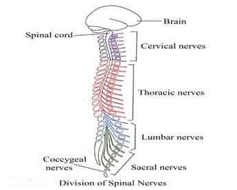
Nerves that arise from the spinal cord are called spinal nerves. There are 31 pairs of spinal nerves in the human peripheral neural system. They include
1. Cervical nerves: Eight pairs
2. Thoracic nerves: Twelve pairs
3. Lumbar nerves: Five pairs
4. Sacral nerves: Five pairs
5. Coccygeal nerves: One pair.
Structure: Each spinal nerve has two types of nerve fibers. These are
1. Afferent nerve fibers (Sensory nerve fibers): They originate from the effecter organ and terminate in the spinal cord
2. Efferent nerve fibers (Motor nerve fibers): They originate from the spinal cord and terminate in effecter organs.
Distribution: Each spinal nerve has two branches.
1. The Posterior branch: It terminates in the muscles and skin of the posterior portion of the body.
2. The Anterior branch: It terminates in the limbs, the lateral and anterior portion of the body.
Certain spinal nerves join to form following plexuses.
1. Cervical plexus: It connects the neck and diaphragm
2. Brachial plexus: It connects chest and arm
3. Lumbar plexus: It connects legs
4. Sacral plexus: It connects the pelvic region
5. Coccygeal plexus: It also connects the pelvic region.
Dr Pramila Singh
