LYMPHATIC SYSTEM
HSBTE HAP DMLT IInd Semester. Lymphatic System: Lymph, Functions of lymph, lymph vessels, lymph capillaries, and lymphoid organs.
Dr Pramila Singh
3/17/20245 min read
HSBTE. HAP. DMLT. IInd-Semester. Lymphatic System: Lymph, Functions of lymph, lymph vessels, lymph capillaries, and lymphoid organs.
Lymph
Lymph is mobile vascular connective tissue made of lymph plasma and lymph cells. Lymph flows inside lymph vessels. Lymph plasma composition is similar to blood plasma. However, lymph plasma has a lower concentration of protein, calcium, and phosphorous than blood plasma. Lymph plasma has a higher concentration of glucose than blood plasma. Lymph cells are mostly WBCs (leucocytes). They float in lymph plasma. RBCs (Erythrocytes) and platelets (Thrombocytes) are not found in the lymph. Thus lymph is a colourless liquid.
Lymph nodes and blood capillaries are sources of leucocytes in the lymph. However, Tonsils, thymus gland, spleen, and Peyer's patch also secret lymph. Lymph drained out in venous blood.
Functions:
1. They carry large macromolecules and large protein molecules into the blood,
2. They carry hormones from endocrine glands to blood,
3. They carry absorbed fat from GIT,
4. They carry the waste product and carbon dioxide into the blood,
5. Lymphocytes from lymph nodes enter into lymph and then into the blood,
6. They carry excess interstitial tissue fluids,
7. They maintain blood volume,
8. They maintain body water volume,
9. They absorb excess water from GIT and slowly release it into the blood
Lymphatic System:
The lymphatic system is made of lymph, lymph vessels, lymph capillaries, and lymphoid organs. Lymph organs include the spleen, bone marrow, thymus, and lymph gland (lymph nodes).
Lymphoid Organs: Rapid formation and maturation of lymphocytes take place in lymphoid organs (lymphatic organs). There are two types of lymphatic organs:
1. Primary Lymphatic Organs (Central lymphatic organs.): Bone marrow and thymus are primary lymphatic organs. T-lymphocyte maturation occurs in the Thymus and B- Lymphocyte maturation occurs in the bone marrow.
2. Secondary Lymphatic organs (Periphery lymphatic organs.): Lymph nodes, tonsils, spleen, Peyer's patches, appendix, etc are secondary lymphatic systems. T- Lymphocytes and B-Lymphocytes enter into secondary lymphatic organs via blood and lymphatic systems. Lymphocytes do not remain in one secondary lymphatic organ for a longer duration. They migrate from one secondary lymphatic organ to another secondary lymphatic organ via blood and lymph.
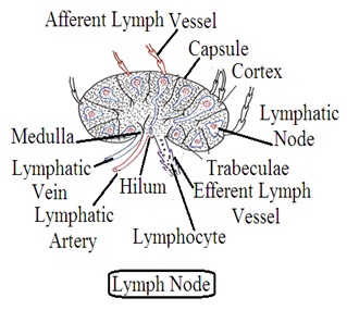
Lymph nodes or Lymph glands: They are bean-shaped. Space inside the lymph node is filled with glandular tissues that secrete lymphocytes (WBCs). They are present in lymphatic vessels and joined with each other by lymphatic vessels. About 400-450 numbers of lymph nodes are present throughout the human body, like in the neck, chest, groin, abdomen, tonsils, etc.
The afferent lymphatic vessel enters into the lymph node through the convex side. Afferent lymphatic vessels pour contents into the space of the lymphatic node. An efferent lymphatic vessel comes out from the hilum side of the lymph node. Lymphocytes along with other material from lymph nodes come out through efferent lymphatic vessels. Blood arteries enter and veins come out from the lymph node through the hilum. The main function of lymph nodes is to produce lymphocytes or WBCs and antibodies. They act as a filter. Lymph node size increases in the area of infection. This swelling may be painful.
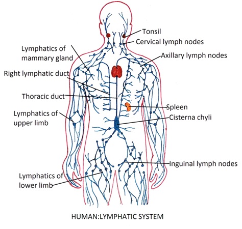
Lymphatic Duct or lymphatic vessel: The lymph duct is connected to lymph capillaries. The lymph duct has several semilunar valves. There are several lymph duct segments between valves. These lymph vessel segments act as an automatic pump. The wall of the lymph duct contracts and pushes lymph to another lymph duct segment in a forwarding direction. Contraction of muscles, body part movement, and tissue compression also help to push lymph into other lymphatic vessel segments. The Union of small lymphatic ducts forms large lymphatic ducts. There are two types of larger lymphatic ducts. These are the thoracic duct and right lymphatic duct.
Dr Pramila Singh


1. Thoracic duct: Lymph from all parts of the body except the right upper part is collected by the thoracic duct. The thoracic duct also collects lymph from the intestine through the lateral lymph duct. This thoracic duct goes upside that receive lymph from the central cervical lymph duct. The left cervical lymph duct carries lymph from the head, neck, chest, and limbs. The thoracic duct pours lymph into a subclavian vein (Brachiocephalic vein),
2. Right lymphatic duct: It carries lymph from the right to the upper part of the body i.e. upper limb, thorax, neck, and head. It is smaller than the thoracic duct. It pours lymph into a right subclavian vein (Brochiocephalic vein). The lymphatic duct swells in an infected body part, the swollen area is painful.
Tonsils: Tonsils are collections of lymphatic tissues present on both sides of the Pharynx. Tonsil mucus is rich in lymphocytes. Tonsils have blood vessels, lymphatic vessels, and lymphocyte mass. These lymphocytes in the tonsils act as the first line of defense. It protects the body against microorganisms that enter the body through the throat, mouth, and nose. In the case of tonsillitis, tonsils cannot protect the body from a microorganism.
Villi: Villi are finger-like the projections in small intestine. It is covered with lymphoid capillaries, blood vessels, and mucus membranes.
Thymus: Epithelial tissues of the pharyngeal wall form the thymus. It is a spherical or oval, soft, pinkish, bilobed mass made of lymphoid tissue. Its size increases up to 15 years of age then its size decreases. It has two lobes connected by connective tissues and enveloped with connective tissue. The outer portion of the thymus is called the cortex and the inner portion is called the medulla. The cortex has a large amount of T-lymphocytes while the medulla has a low amount of T-lymphocytes.
Reticulo Endothelial System (RES): Reticuloendothelial system is a specialized system to perform phagocytosis. Their main function is to protect the body from infectious microorganisms and toxins. They are mainly found in lymph glands, bone marrow, and liver.
Spleen: Spleen is present on the left side of the abdomen in between the stomach fundus and diaphragm. Its outer surface is in contact with the diaphragm. It touches the colon, left kidney, and pancreas tail. It is protected by the ninth, tenth, and eleventh ribs. It is soft, dark purple, and rich in vascular supply. It is the largest lymphoid organ. Splenic blood vessels enter the spleen through the hilum and exit from the spleen through this hilum.
There is no blood capillary inside the spleen. Thus blood remains in direct contact with spleen cells. The artery of the spleen is called the splenic artery & the vein is called the splenic vein.
Functions: It produces all types of blood cells in infants, Lymphocytes in adults, and RBCs in the adult during weakened functions of bone marrow. Both B-lymphocytes & T-lymphocytes multiply in the spleen.
B- cells: In humoural immune system
T-cells: Cells mediated immune system
B- cells: Synthesised in bone marrow & liver
T-cells: Synthesised in thymus gland
B- cells: Acts against pathogens, no action against cancer cells, and transplant
T-cells: Acts against pathogens, cancer cells, and transplant
B- cells: Release antibodies to destroy antigens
T-cells: Attack directly on antigens
B- cells: Secretes antibodies into the blood
T-cells: Does not secrete antibody
