Human Eyes
Human Eyes Structure, Functions, and Defects
Dr Pramila Singh
2/24/20248 min read

HSBTE, DMLT, IInd Semester, HAP. Human Eye: Structure, Functions and Defects
ORGAN OF SIGHT (Eyes)
Human eyes act as a portal to collect data from surroundings and forward them to the brain for processing.
Structure of eyes:
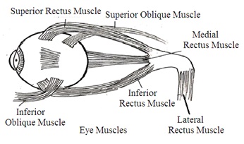
The human body has two eyes as an organ of sight. They are located in the orbit of the skull (eye socket or eye orbit). Each Eye has a round structure called an eyeball. Eye orbit has a fat cushion that protects the eyeball from jerking and helps in frictionless rotation or movement of the eyeball. Both eyeballs move simultaneously. Four rectus muscles (straight muscle) and two oblique muscles hold and move the eyeball inside the eye orbit.
A. Rectus muscles (Straight muscle) are responsible for eyeball movement as per below mentioned directions:
1. Superior rectus muscle: Upward movement of the eyeball,
2. Inferior rectus muscle: Downward movement of the eyeball,
3. Medial rectus muscle: Inward movement of the eyeball,
4. Lateral rectus muscle: Outward movement of the eyeball.
B. Oblique muscles are responsible for eyeball movement as per below mentioned direction
1. Superior oblique muscle: Internal rotation of the eyeball that is looking towards the nose, looking away from the nose, and looking downward.
2. Inferior oblique muscle: Upward & outward eyeball movement, external rotation, elevation, and adduction of the eyeball.

Eyeball
The eyeball is a hollow oval structure made of a three-layered wall. These layers are
1. Outer fibrous layer (Fibrous Coat): Supporting part of the eyeball, Sclera, and Cornea.
2. Middle Vascular layer (Vascular Coat): Choroid, iris, and Ciliary body
3. Inner nervous layer (Nervous Coat): Retina.
OUTER FIBROUS COAT (Fibrous Tunic or Fibrous Layer or Fibrous Part): It is the outer layer of the eyeball. It is the toughest layer and consists of fibrous elastic connective tissues without any nervous tissue. It is a Supporting part of the eyeball. There are two parts in a fibrous coat: Sclera and Cornea.
i. The sclera is made of tough fibrous connective tissue with collagen fibers. It is the opaque white part of the eyeball covering about 60% of the posterior portion of the eyeball. The sclera provides a surface for muscle attachment. A very small portion of the sclera is visible from the front of the eyeball. Most of its part is present inside the skull orbit and not visible. It gives shape to the eyeball and protects the eyeball.
ii. The cornea is an anterior (front) thick transparent portion of the eyeball. It covers about 40% of the anterior eyeball fibrous coat. Most of its portion is visible from the front side of the eyeball. The cornea is a non-vascular (no blood supply) part of the eyeball. The cornea gets its nourishment from the aqueous humor, lachrymal secretion, and lymph. The cornea can absorb oxygen from the air.
The canal of Schlemm is present at the junction of the cornea and sclera. It is called Sclerocorneal Junction. It drains out excess aqueous humor from the eyeball anterior chamber into blood veins.
The outer layer of the cornea and the exposed portion of the sclera are covered by a transparent membrane called the conjunctiva.
VASCULAR COAT (Uvea): It is a middle vascular pigmented layer of eyeball present under the sclera. It is made of three parts iris, ciliary, and choroid.
1. The choroid is the most vascular part of the vascular coat (uvea) present just under the sclera. It supplies oxygen and nutrients to another part of the eyeball. It has maximum pigmented cells. Usually, the choroid is dark brown due to the presence of melanin pigment.
2. Ciliary Body: It is a vascular part present in between the iris and choroid. It is made of less pigmented ciliary smooth muscle and ciliary processes. Ciliary muscles act as sphincters and control the iris.
The ciliary body is attached to the eye lens capsule by suspensory ligaments. The ciliary body, suspensory ligaments, and eye lens capsule hold the eye lens. They help in the accommodation of eyes. Ciliary processes epithelium secretes aqueous humor.
3. Iris: Iris is a pigmented, circular, muscular perforated diaphragm. It starts from the ciliary body and is present in front of the eye lens. The Central perforated part of the iris is called the pupil. Iris has Circular smooth muscles and radial smooth muscles that control the size of the pupil. Contraction in circular smooth muscles causes constriction of the eye pupil. Contraction in radial smooth muscle causes dilation of the pupil.
These smooth muscles control the entry of light into the eye chamber. In bright light, Circular muscles constrict to decrease the size of the pupil (Miosis). It restricts the entry of light into the eye chamber. In dim light, dilator muscles (Radial muscles) constrict to increase the size of the pupil (Mydriasis). It allows the entry of more light in the dark.
The Colour of the eye depends upon the melanin (pigment) present in the iris. A small amount of melanin in the iris develops green-colored eyes. Lack of melanin in iris develops blue coloured eye. Lots of melanin in iris develops a brown-coloured eye.
NERVOUS COAT: It is the third innermost nonvascular sensory layer of the eyeball wall. It is called the Retina. The retina consists of the following two layers:
i. The outer surface of the retina is present in contact with the choroid. It consists of a single layer of pigment cells.
ii. The inner layer of the retina is present in contact with Vitreous fluid. The inner layer of the retina has a yellow area called Macula lutea.
· The middle of the macula lutea has a small shallow depression called Fovea centralis (Yellow spot). A yellow spot is present just opposite the cornea. It has a maximum number of cone cells and no rod cells. This is a place of sharpest vision.
· Optic nerve fibers and blood vessels enter into and come out from the eyeball through a Fovea centralis (yellow spot) in the retina.
· Macula lutea has a blind spot with no receptor cells. There is no image formation at the blind spot.
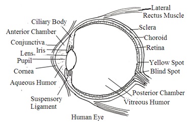

OTHER PARTS OF THE EYEBALL: Eyeball has also the following parts: Lens, Aqueous humor, and Vitreous humor.
1. Lens: The lens is a biconvex elastic body present just behind the iris (Behind the pupil). Suspensory ligaments hold the lens inside the eyeball and connect it with the ciliary body. The lens is covered with a thin transparent proteinous membrane called a capsule (Lens capsule). Change in the curvature of the lens helps to focus light rays on the retina. The ability to change lens curvature by contraction and relaxation of suspensory ligaments and ciliary body is called the Power of Accommodation. Eye lens, suspensory ligaments, and ciliary muscles divide the eyeball chamber into two parts: The aqueous chamber and the Vitreous chamber.
2. Aqueous chamber: It is filled with aqueous humor. The aqueous chamber is further divided by iris into two parts anterior aqueous chamber and posterior aqueous chamber.
· The anterior chamber is in front of the iris.
· The posterior chamber is behind the iris. These two chambers are interconnected through the eye pupil.
Aqueous humour: It is a transparent aqueous fluid. It develops pressure in the aqueous chamber to maintain the proper shape of the eyeball front part. It also provides nutrition to the eye lens and cornea. The canal of Schlemm continuously drains out aqueous humour from the aqueous chamber.
3. Vitreous chamber. The chamber between the lens and the retina is the vitreous chamber. It is filled with vitreous humour. Vitreous Humour is a transparent jelly-like substance. It maintains the shape of the eyeball. It is non-replaceable, does not flow, and is drained out.
ACCESSORIES of EYES (Appendages of the eyes):
Accessories of the eyes are eyebrows, eyelids, eyelashes, conjunctiva, and lachrymal apparatus. They mainly protect the eyes.
1. Eyebrows: An eyebrow is an arch of thick skin just above each eye. It has numerous hairs. Eyebrows protect the eyeball from dust, foreign bodies, sweat, and light.
2. Eyelids: Above and below the eye front has movable folds called eyelids. They are covered by the skin at the upper surface and conjunctiva at the lower surface. The upper eyelid is bigger than the lower eyelid. The free edges of both eyelids have hair called eyelashes. Eyelashes protect the eyeball front part from light and dust. The edge of the eyelid produces oily lubricant to lubricate the cornea of the eyes.
3. Conjunctiva: It is a transparent mucus membrane present on the inner surface of eyelids that covers the front of the cornea and sclera of the eyeball.
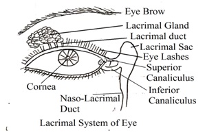
4. Lacrimal apparatus: It consists of the lacrimal gland (tear gland) and various ducts. Lacrimal glands are almond-shaped glands that secrete tears. Tears are watery fluids consisting of water, salt, sugar, amino acids, lysozyme, and mucus secretion. Lysozymes are bacteriocidal proteinous enzymes that kill bacteria. Tears from the lacrimal gland may enter the nasal cavity through the nasolacrimal duct.
Tears moisten the anterior part of the eyeballs and the inner surface of the eyelids. It washes out dust, dirt, and microbes present on eyeballs and the inner surface of eyelids. It also nourishes the cornea. Protection, moistening, and cleaning of the eye occur during the normal blinking of eyelids.
5. Adipose tissues: Adipose tissues are present inside the eye orbit around the eyeball. They act as a cushion to make eyeball shockproof.

WORKING OF EYES (Mechanism of vision): Eyes receive light rays stimuli on the retina and pass sensory impulses to the brain's visual center through optic nerves.
1. Image formation on the retina: Light rays from the object enter into the eyeball through the conjunctiva, cornea, aqueous humour, pupil, lens, and vitreous humour.
· Iris's muscles regulate light rays entry into the eyeball by increasing or decreasing the size of the pupil.
· Iris's muscles also change lens curvature to focus the image on the retina fovea centralis (Yellow spot).
· Lens forms a small and inverted object image on the retina
· Cornea forma is a rough image and the lens forms a fine image on the fovea centralis (Yellow spot) of the retina.
2. Power of accommodation: The ability of the eye to change lens curvature to focus object image on the retina is called the power of accommodation (Accommodation reflex).
· The eye can view objects up to a distance of 25 cm. Objects at a distance of less than 25 cm are not visible. This 25 cm distance is considered as near point. At a near point, eyes show maximum accommodation with the lens at maximum curvature (convexity).
· The image of the object at a far distance falls on the retina without any accommodation in the curvature of the lens. Eyes do not require the power of accommodation to view objects at a distance of 6 meters or more.
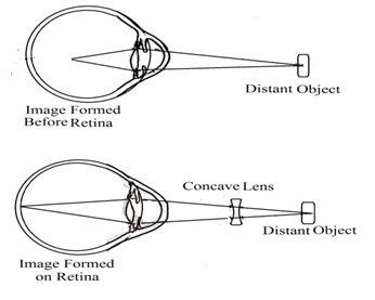


Eyes Defects: Myopia (Short-sightedness or near-sightedness): The lens forms an image of distal objects in front of the retina, not on the retina. Near objects will be visible but distal objects will be blurred in vision.
Myopia can be solved by using concave lens spectacles.
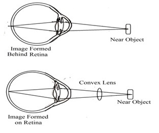
Hypermetropia (Far far-sightedness or Long-sightedness): It forms an image of near vision behind the retina, not on the retina. It makes closure objects blurred vision, while far away objects will be visible clearly.
Vision can be corrected by using a convex lens.
Conjunctivitis: Inflammation in the conjunctiva is called conjunctivitis. It may be acute or chronic. Symptoms are photophobia, redness in the conjunctiva, wasting of eyes, swelling in the eyelid, filling of heat and grittiness in eyes.
