
HSBTE Hematology
HSBTE Question Paper Hematology, Ist Semester, Examination July 23. Questions with Solution
HSBTE QUESTION SOLUTION
Alok Bains
12/10/20239 min read
HSBTE Question Paper Hematology, Ist Semester, Examination July 23. Questions with Solution
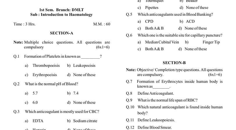
1st Sem. Branch: DMLT. Subject Code: 221916
Subject: Introduction to Haematology, Time : 3 Hrs. M. M.: 60
SECTION-A
Note: Multiple choice questions. All questions are compulsory (6x1=6)
Q.1 Formation of Platelets in known as _________?
a) Thrombopoeisis b) Leukopoeisis c) Erythropoeisis d) None of these.
Ans: a) Thrombopoeisis
Q.2 What is the normal pH of Blood?
a) 5.7 b) 7.4 c) 6.0 d) None of these
Ans: b) 7.4
Q.3 Which anticoagulant is mostly used for CBC?
a) EDTA b) Sodium citrate c) Heparin d) None of these
Ans: a) EDTA
Note: EDTA: Ethylenediamine Tetraacetic Acid. CBC: Complete Blood Coun
Q. 4 Which one is used during venous Blood collection?
a) Tourniquet b) Beaker c) Pipettes d) None of these
Ans: a) Tourniquet
Q.5 Which anticoagulants are used in Blood Banking?
a) CPD b) ACD c) Both A & B d) None of thes
Ans: c) Both A & B
Note: CPD: Citrate Phosphate Dextrose (in solution form). ACD: Acid Citrate Dextrose (in solution form).
Q.6 Which one is the suitable site for capillary puncture?
a) Median Cubital Vein b) Finger Tip c) Both A & B d) None of these
Ans: b) Finger Tip
Note: The middle or Ring fingertip of the non-dominant hand is used for capillary puncture because they are less sensitive to pain. They are also less calloused than the thumb and index finger. The thumb is also avoided due to the presence of more arteries in the thumb
SECTION-B
Note: Objective/ Completion type questions. All questions are compulsory. (6x1=6)
Q.7 Formation of Erythrocytes inside the human body is known as________.
Ans: Erythropoiesis.
Note: Erythropoiesis occurs in the bone marrow.
Q.8 Define Anticoagulant.
Ans: An anticoagulant is a substance that prevents blood from clotting or reduces the formation of blood clots. Examples are Heparin, EDTA, CPD, ACD, etc. Heparin is a natural coagulant present in human blood inside the human body's blood Vessels.
Q.9 What is the normal life span of RBC
Ans: 120 days.
Q.10 Which natural anticoagulant is found inside the human body?
Ans: Hepari
Q.11 Define Leukoopoiesis
Ans: The process of white blood cell (leukocyte) formation or production in the body is called Leukopoiesis.
Note: Leukopoiesis is a part of hematopoiesis. The overall process of forming blood cells is called hematopoiesis
Q.12 Define Blood Smear.
Ans: Blood smear is a laboratory technique to form a thin layer of blood on a microscope slide.
Note: A blood smear is also known as a peripheral blood smear or peripheral blood film. The blood smear provides detailed information about the cellular components of blood, including red blood cells (erythrocytes), white blood cells (leukocytes), and platelets.
SECTION-C
Note: Short answer type questions. Attempt any eight questions out of ten questions. (8x4=32)
Q.13 Write the various functions of blood.
Ans: Blood is composed of plasma and blood cells. Each component performs its functions.
A. Plasma Proteins Functions: Blood plasma has several proteins. These proteins transport absorbed food, respiratory gases, metabolic wastes, hormones, antibodies, heat, clotting factors, etc.
B. Function of Red Blood Cells (Erythrocytes): RBC contains hemoglobin that transports respiratory gases and maintains blood pH.
C. Function of White Blood Cells (Leucocytes): Protection of the body from microorganisms and toxins produced by the microorganism.
D. Function of blood platelets: They help in blood coagulation and clot formation.
Q.14 Draw and write the uses of Hb. RBC and WBC Pipette.
Ans: Hemoglobin (Hb) pipette, Red Blood Cell (RBC) pipette, and White Blood Cell (WBC) pipette are laboratory instruments used in hematology for the measurement and analysis of blood components.
A. Hemoglobin (Hb) Pipette
Hemoglobin Pipette is used for the collection of blood from capillary puncture for hemoglobin estimation. It has a single mark of 20 cu mm.
B. Red Blood Cell (RBC) Pipette
C. The RBC pipette is a diluting pipette. The RBC pipette is used to dilute blood samples for RBC count in the Hemocytometer. RBC pipette has a red bead in the bulb and it has markings of 0.5, 1, and 101. Red bead helps in the mixing of diluting fluid and blood.
D. White Blood Cell (WBC) Pipette
WBC pipette is a diluting pipette. The WBC pipette is used to dilute blood sample for RWBC count in the Hemocytometer.
Q.15 Define Plasma and write their compositions.
Ans: Definition: Blood plasma is the yellowish liquid component of blood that contains soluble components of blood and suspended blood cells. Blood plasma constitutes 55% of the total blood volume.
Composition of blood plasma: It consists of the following componenets
A. Water: Approximately 90-92% of blood plasma volume is water.
B. Proteins: Albumin, Globulin, Fibrinogen, etc.
C. Electrolytes: Sodium, Potassium, Calcium, Chloride, etc.
D. Gases: Oxygen, carbon dioxide, etc.
E. Nutrient: Glucose, amino acids, fatty acids, etc.
F. Hormones,
G. Waste Products: Urea, Creatinine, and other metabolic wastes.
Q.16 Describe Non-Calcium chelators anticoagulants.
Ans: Anticoagulants are substances that prevent blood from clotting. Non-calcium chelators and anticoagulants act independently without involving calcium ions in the blood. The following two are commonly used non-chelator anticoagulants
1. Heparin: Heparin is a natural anticoagulant that combines with antithrombin III and catalyzes antithrombin-III ability to inactivate thrombin and other clotting factors.
2. Argatroban: It is a direct thrombin inhibitor. It does not require antithrombin III for its anticoagulant effect
Q17 Describe the selection and preparing of the vein puncture site.
Ans: SELECTION AND PREPARING OF VENIPUNCTURE SITE
Selection of site for venipuncture: The following veins are suitable for venipuncture.
Inside antecubital fossa located in the elbow
a. Cephalic vein: The cephalic vein runs an entire arm's length.
b. Basilic vein: A basilic vein is a vein located in the triangular area inside the elbow. The basilic vein is considered an alternate site for venipuncture. It is used if other more prominent arm vein is not visible.
c. Median cubital vein: The median cubital vein connects the cephalic vein and basilic vein. It is preferred due to its large size and lower tendency to roll during needle insertion. Very few nerve endings surround this vein. Thus it is less painful to insert the needle into a median cubital vein.
These three veins are superficially located in the triangular area inside the elbow.
d. Superficial veins in foot and ankle
The median cubital vein and cephalic vein near the median cubital vein are the most common and suitable sites for vein puncture. Sometimes hand vein is selected if these three veins are not prominent. Hand veins tend to roll. Thus the skin is pulled taut then a needle is inserted. Superficial veins in the foot and ankle are considered the last alternate site for venipuncture. However, the following sites (areas) should be avoided for venipuncture.
a. Site having scars due to burning or surgery. It is difficult to puncture scar tissue.
b. Mastectomy site. This area has lymphodema. That affects the blood sample test result.
c. Bad bruise (Haematoma) on the skin,
d. Skin part used for intravenous therapy or blood transfusion.
e. Skin with an infection like eczema or any other disease condition.
f. Arm with canula or fistula or heparin leak. This site should be used only after physician consultation.
g. Endematous extremities. This area has tissue fluid accumulation. These fluids may enter blood samples. That will dilute blood and affect blood test reports.
h.Thrombosed vein.
Preparation for venipuncture: Select the venipuncture exact site by tracing the palpating vein using the index finger. If palpitation is not felt easily then the following measures are taken to improve blood flow towards the venipuncture site
i. The arm is massaged from the wrist to the triangular inner side of the elbow.
ii. Tap the venipuncture site by using the index finger and middle finger.
iii. Apply a warm moist cloth to the venipuncture site at least for 5 minutes,
iv. Lowering of the arm below bed level also allows the vein to be filled with venous blood.
Q.18 Write the materials and equipment required for capillary puncture.
Ans: MATERIALS AND EQUIPMENTS REQUIRED FOR CAPILLARY PUNCTURE SITE:
Capillary punctures require different devices such as lancets, microcontainer tubes, microhematocrit tubes, sealants, alcohol (ethyl alcohol or isopropyl) as a disinfectant, and warming devices. Improper use of these devices may cause improper specimen collection and pre-analytical errors.
i. Lancets are meant to puncture or cut the skin to collect capillary blood. They are specifically designed for finger or heel puncture.
ii. Microcontainer tubes is used to collect minute volumes of blood via capillary puncture. They are coated with different additives.Colour code on microcontainer tube represents the additive present inside it. They have also markings indicating the volume in µL.
iii. Microhematocrit tubes are glass or plastic capillary tubes. They are used for blood collection and hematocrit determination. They are either coated with anticoagulant or plain. Plastic or clay sealants are commonly used. It makes Microhematocrit tubes leak-proof seal.
iv. Warming device (heel warmer): It is used to increase the arterial blood flow at the puncture site. It makes a close resemblance of sample blood with arterial blood to venous blood. A towel dampened with warm water can be used to produce the same effect as a warming device. However, water should not be too hot to burn the patient.
Q.19 Describe the Vacutainer system in brief.
Ans: VACUTAINER SYSTEM: It is also known as an evacuated collection tube. It is made up of glass or plastic. If an additive is present in the collection tube then it is called an additive collection tube. If the collection tube has no additive then it is called a non-additive collection tube. The collection tube has a colored rubber or plastic top (colored rubber or plastic stopper). This color is used to indicate the specific hematological test to be performed by using blood present in the collection tube. This color also indicates the additives added to the collection tube.
A sterilized collection tube (vacutainer) is used to collect and store blood from the vein of the patient. The sterilized top of the collection tube is inserted into a connector attached to a butterfly needle. The tube is further pushed into the connector. This creates a vacuum in a sterilized collection tube. The top of the needle is pierced into a vein of the patient. Blood automatically enters into a sterilized collection tube due to the vacuum created by the collection tube.
A collection tube (vacutainer) is a closed system to withdraw blood from a vein through a venipuncture procedure. This prevents exposure of blood to air or outside contaminants. It is available in various sizes and volumes. This size is selected as per requirements such as the age of the patient, volume of blood to be collected, condition of the patient’s vein, and size of the patient’s vein.
Each collection tube has an expiry date that is for the additive and vacuum system of the collection tube. The collection tube should be filled properly. Improper filling shall disturb blood and additive proportion (ratio). This will not give accurate test results. This type of sample shall also be rejected by a laboratory technician.
Q.20 Write the procedure of preparation of Thick blood film.
Ans: A thick blood film is commonly used in the diagnosis of malaria and other bloodborne parasites.
Procedure:
1. Prepare the Patient: Explain the procedure to the patient and obtain informed consent. Select a suitable site for blood collection (commonly the fingertip or earlobe), Clean the selected site with an alcohol swab.
2. Collect Blood: Use a lancet or needle to puncture the cleaned site and collect a small drop of blood on one end of the clean microscopic slide.
3. Make a Thick Blood Smear: Spread the blood in a circular motion to create a thick and even film by using a spreader slide. The size of the smear should be about the diameter of a dime or slightly larger.
4. Allow the Blood Smear to Air-Dry: Allow the thick blood smear to air-dry completely. This can take several minutes.
5. Fix the Blood Smear: Fix the blood smear by immersing the slide in methanol or other fixative for 1-2 minutes. This step helps in preserving the cellular morphology.
6. Rinse and Dry: Rinse the stained slide with distilled water to remove excess stain. Allow the slide to air-dry completely.
Q.21 Describe the Romanowsky stain in brief.
Ans: Romanowsky stains are a group of histological stains used in microscopy to color and differentiate various components of cells. These stains are commonly employed in hematology for the microscopic examination of blood cells and blood-borne parasites. One of the most well-known Romanowsky stains is Giemsa stain, which is widely used for staining blood smears in the diagnosis of malaria and other blood-related disorders,
Romanowsky stain consists of water-soluble eosin, methylene blue, and acetone-free methanol.
1. Eosin is an anionic acidic dye that imparts pink and red color to cellular structure. Eosin combines with cationic components of cells eg Cytoplasm.
2. Methylene blue is a cationic basic dye that imparts blue-purple colour to cellular structure. Methylene blue combines with an anionic component of cells eg DNA.
3. Acetone is a strong decolourising agent and a strong dehydrating agent. Thus acetone-free methanol (Absolute methanol) is used as an ingredient in the Romanowsky stain. This acetone will destroy the cell membrane. Methanol acts as a fixative.
Pink-red or deep red color Red blood cells.
Gray-blue color: Reticulocytes.
Pale pink cytoplasm, purple granule Neutrophil
Pale pink cytoplasm, Eosinophil
Blue cytoplasm, Dark blue Basophil
Blue cytoplasm Monocytes
Dark blue cytoplasm Lymphocyte
Purple color Platelets
Bluish purple RNA, DNA.
Q.22 Describe Thrombopoiesis in brief.
Ans: Thrombopoiesis is the process of platelets (thrombocytes) formation. It is a part of hematopoiesis (the formation of blood cells). Thrombopoiesis occurs in the bone marrow. It involves a series of steps to produce mature platelets.
1. Hematopoietic Stem Cells: Thrombopoiesis begins with hematopoietic stem cells (HSCs) in the red bone marrow. Megakaryocytes of stem cells develop platelets. One megakaryocyte develops 1000 platelets. Hormone thrombopoietin stimulates the production of platelets.
2. Megakaryocyte Maturation: Megakaryocytes are large, multinucleated cells. They undergo maturation within the red bone marrow. They develop a complex cytoplasmic structure with multiple lobes. These are mature Megakaryocytes (Proplatelets).
3. These mature Megakaryocytes undergo fragmentation and form small platelets.
4. The newly formed platelets are released into the bloodstream from the bone marrow.
5. Platelets Lifespan: Platelets have a relatively short lifespan about 7-10 days.
SECTION-D
Note: Long answer type questions. Attempt any two questions out of three questions. (2x8=16)
Q.23 Describe the development stages of the Erythrocyte in detail.
Q.24 Explain various anticoagulant vials/tubes with their color code and uses.
Q.25 Explain the Requirement, Preparation, and procedure for capillary blood collection.
Dr Pramila Singh