Giardia
Giardia Morphology and Life cycle
PARASITOLOGY
Dr Pramila Singh
10/12/20233 min read


Morphology of Giardia
Giardia is a pear-shaped single-celled parasite. It exists as a trophozoite and cyst.
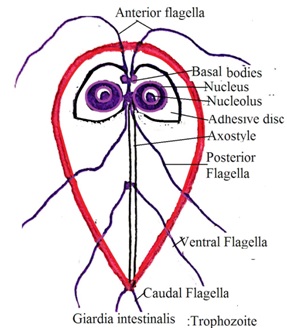
A. Morphology of Giardia Trophozoite:
· It is an active motile form of Giardia. It has a flat bilateral symmetrical body.
· Its length varies from 10 to 20 micrometers and width 5 to 15 micrometers.
· Its ventral surface is concave and the dorsal surface is convex. The ventral surface has pair of adhesive discs (sucking discs) towards the anterior end. The dorsal surface has an axostyle. These structures attach the Giardia trophozoite to the intestinal wall of animals and also help in locomotion.
· It has a broader anterior end and a pointed posterior end.
· It is a bi-nucleated parasite. It has two nuclei near the anterior end. Both nuclei are connected by microtubules called median axoneme or median body.
· It has four flagella four on each side (Four pairs of flagella): Two from the lateral side, two from the middle, two from the anterior, and two from the posterior end of the parasite.
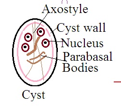
B. Morphology of Giardia Cyst:
Giardia cyst is produced from the Giardia trophozoites. Giardia cyst is a dormant, ineffective form of the Giardia.
a. It is oval-shaped with measurements of about 8 to 12 micrometers.
b. It is covered by a protective layer to protect Giardia from unfavorable conditions such as changes in temperature and pH.
c. Its outer surface has a dividing line called axostyle.
Inside the Giardia cyst, the Prabasal body exists.
d. It has two to four nuclei and other cellular structures.
e. Sucking disc points and flagella points are visible in the cytoplasm of the Giardia cyst. It comes out from the infected individual or animal body with stool.
f. It can survive up to one month in the atmosphere outside the human body or animal.


Life cycle of Giardia
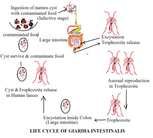
Giardia is a microscopic parasite that causes diarrhoea in humans. This diarrhoea is called giardiasis. There are two stages in the life cycle of the Giardia: The Trophozoite stage and the Cyst stage.
1. Trophozoite stage: Trophozoites are active, motile forms of the Giardia. It enters into host body with infected water or food. The host stomach has an acidic pH. Acidic media is unfavourable for Giardia trophozoite. Thus, it enters the host intestine quickly. It is attached to the infected host intestine. One Giardia trophozoite produces two trophozoites by asexual reproduction inside the intestine of the infected host.
2. Cyst stage: Inside the large intestine, Giardia trophozoite develops a thick wall around it to form a cyst. ThIs cyst has two Giardia cells, Trophozoite undergoes cell division to form two cells of Giardia. These cysts are excreted out from the host body with stool. Some trophozoites are also excreted with stools. However, these trophozoites do not survive outside the human body. Giardia cysts contaminate soil, water and food.
Transmission of infection: Ingestion of food contaminated with Giardia cyst develops giardiasis in the infected host. Inside the duodenum and jejunum of the host, it develops Giardia trophozotes.


Lab diagnosis of Giardia
There are several methods to detect and identify Giardia parasites. Administration of anthelmintic drugs at bedtime and collection of stool samples in the morning is the most suitable technique to detect and identify parasites. The following are commonly used techniques to detect and identify Giardia parasites
1. Microscopic examination: Microscopic examination of stool samples is a primary method to detect and identify Giardia parasites. Fresh and concentrated sample of stool is examined under a microscope. Both Giardia trophozoite and Giardia cyst are present in the infected stool sample.
Giardia trophozoite is identified by its characteristic pear-shaped structure and its characteristic motility. Characteristic motility is “falling-leaf” or “tumbling’ motility.
2. Stool antigen detection: Stool infected with Giardia has a specific Giardia antigen. Giardia antigen is detected by enzyme immune assays (EIAs) or immunochromatographic assays (ICAs). It provides rapid results.
3. Stool PCR (Polymerase Chain Reaction): PCR test is used to detect and identify Giardia DNA in stool samples. It is a highly sensitive and specific method to differentiate various species of Giardia. It is especially useful to detect Giardia when a low concentration of Giardia is in stool samples. It works even though other laboratory diagnostic tests fail to detect and identify Giardia in stool samples.
4. Stool culture: It is a less commonly used technique to detect and identify Giardia in the sample. A stool sample is inoculated in the suitable culture media. Inoculated culture media is incubated. Trophozoites are identified microscopically.
5. Immunofluorescence: Fluorescent antibodies are used to stain Giardia.Fluor4escent microscope is used to detect and identify Giardia trophozoite and cyst.
6. Clinical diagnosis: Yellow-grey, foul-smelling without blood diarrhoeal stool is a clinical symptom of Giardiasis.
Dr Pramila Singh
