ESR and PCV Estimation
UNIT I ESR and PCV Estimation 1.1 Erythrocyte sedimentation rate (ESR) and packed cell volume (PCV) Introduction 1.2 Various methods of estimation of ESR and PCV. 1.3 Merits and demerits during the estimation of ESR and PCV. 1.4 Factors involved in ESR estimation. 1.5 Interpretation of results. HSBTE, IIIrd Semester DMLT
HAEMATOLOGY
Dr Pramila Singh
9/4/20236 min read
UNIT I
ESR and PCV Estimation: Erythrocyte sedimentation rate (ESR) and packed cell volume (PCV), Introduction, Various methods of estimation of ESR and PCV. Merits and demerits during the estimation of ESR and PCV. Factors involved in ESR estimation. Interpretation of results. HSBTE, IIIrd Semester DMLT
1.1 Erythrocyte sedimentation rate (ESR)
Erythrocyte sedimentation rate (ESR) is a blood test to measure the rate of sedimentation of red blood cells in a long tube over a specific period of time. It is a useful test to detect and monitor inflammatory conditions. ESR increases during inflammatory conditions in the human body. The body produces fibrinogen and C-reactive protein (CRP) in the blood during any inflammatory conditions. These proteins cause clumping in the RBCs. RBCs become heavier which increases their rate of sedimentation.
An increase in ESR occurs in various conditions such as infections, auto-immune diseases, cancers, tissue damage, etc. Only ESR is not used to diagnose disease. The disease must be confirmed by using other diagnostic tests.
Packed cell volume (PCV)
Packed cell volume (PCV) is also called hematocrit. PCV is a blood test to measure the volume of red blood cells or percentage of red blood cells in a given sample of blood. It is measured to know the oxygen-carrying capacity of the blood and to evaluate anemia, dehydration, polycythemia, and other conditions related to blood volume. PCV tests with other blood tests are used to diagnose blood conditions.
1.2 Various methods of estimation of ESR and PCV
Methods of estimation of ESR: There are two commonly used methods to measure ESR. These are the Westergren method and the Wintrobe method.
Westergren method for ESR estimation: It is the most widely and most preferred method to measure ESR.
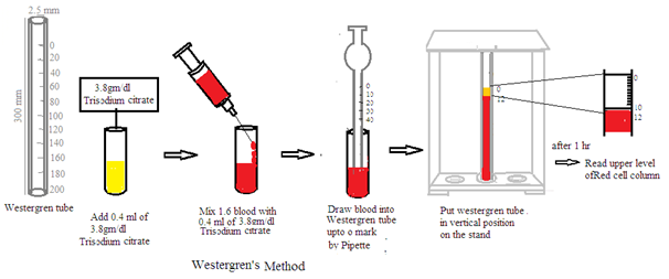
Equipment: The Westergren method requires a Westergren tube. It is a long narrow graduated glass tube 300 mm long and 2.5 mm diameter. It is marked 0 to 200 mm. It looks like a 1 mm pipette. It holds 2ml of blood.
Anticoagulant: A 3.8% trisodium citrate solution is used as an anticoagulant in ESR measurement by the Westergren method. 4:1 Blood: anticoagulant ratio is used to stop blood clotting during the ESR test.
Blood sample: Collect the patient's venous blood. Use the blood sample within 2 hours. Blood samples can be preserved at 4 degrees C. But preserved blood sample should be used within 6 hours.
Dilution: Mix 1.6 ml of blood with 0.4 ml of 3.8% trisodium citrate solution thoroughly and gently.
Filling the tube: Use a pipette to draw blood samples into the Westergren tube. Avoid mouth suction of blood sample. Draw diluted blood sample into Westergren tube until blood level reaches up to zero marks on the tube.
Placing the Westergren tube: Press the top of the Westergren tube. Set the filled Westergren tube vertically in a Westergren stand. Allow the blood to settle for 60 minutes without disturbing.
Reading result: After 60 minutes, measure the distance in mm between the top of the clear plasma and red blood cells column. This is ESR mm/hr.
The normal range in males is 4 to 14 mm/ 1 hr and in females 6 to 20 mm/1 hr.
Interpretation: ESR value is reported in mm/hr. Normal ESR values vary with age and gender. It is a non-specific test. Inflammation, infection, auto-immune disease, cancer, and certain medical conditions increase the ESR value. Polycythaemia, Congestive cardiac failure, anemia, etc. decrease ESR.
Wintrobe method to estimate ESR value:
The Wintrobe method is another most commonly used method to estimate ESR.
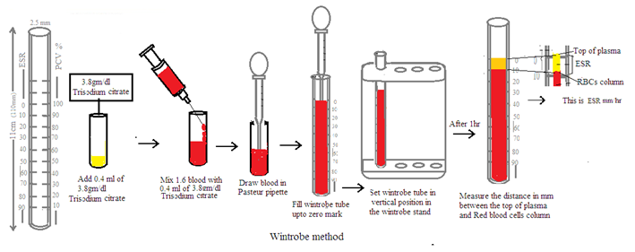
Equipment: Wintrobe tube is a long narrow and graduated glass tube 11cm long, 2.5mm internal diameter, and flat inner base. It is marked zero to 100 in a reversed direction. Descending marking is used to estimate ESR. Ascending marking is used to estimate PCV. It holds 1 ml of blood.
Anticoagulant: A 3.8% trisodium citrate solution is used as an anticoagulant in ESR measurement by the Wintrobe method. 4:1 Blood: anticoagulant ratio is used to stop blood clotting during the ESR test.
Blood sample: Collect the patient's venous blood. Use the blood sample within 2 hours. Blood samples can be preserved at 4 degrees C. But preserved blood sample should be used within 6 hours.
Dilution: Mix 1.6 ml of blood with 0.4 ml of 3.8% trisodium citrate solution thoroughly and gently.
Filling the tube: Use a pipette to draw blood samples into the Wintrobe tube. Avoid mouth suction of blood sample. Draw diluted blood sample into Wintrobe tube until blood level reaches up to zero marks on the tube.
Placing the Wintrobe tube: Press the top of the Wintrobe tube. Set the filled Wintrobe tube vertically in a Wintrobe stand. Allow the blood to settle for 60 minutes without disturbing.
Reading result: After 60 minutes, measure the distance in mm between the top of the clear plasma and red blood cells column. This is ESR mm/hr.
The normal range in males is 0 to 9 mm/ 1 hr and in females 2 to 20 mm/ 1 hr.
Interpretation: ESR value is reported in mm/hr. Normal ESR values vary with age and gender. It is a non-specific test. Inflammation, infection, auto-immune disease, cancer, and certain medical conditions increase the ESR value. Polycythaemia, Congestive cardiac failure, anemia, etc. decrease ESR.


Methods of Estimation of PCV
There are two methods to estimate PCV (Haematocrit). These are the macro method using a Wintrobe tube and the micro method using a capillary tube.
Macromethod using Wintrobe tube: It is the Wintrobe method to estimate PCV in the blood sample. The following steps are followed in the Wintrobe method
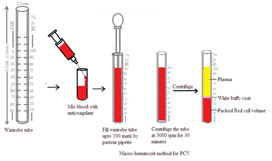
Equipment: The Wintrobe tube is a long-narrow and graduated glass tube 11cm long, 2.5mm internal diameter, and a flat inner base. It is marked zero to 100 in a reversed direction. Descending marking is used to estimate ESR. Ascending marking is used to estimate PCV. It holds 1 ml of blood.
Anticoagulant: A 3.8% trisodium citrate solution is used as an anticoagulant in ESR measurement by a Wintrobe method. 4:1 Blood: anticoagulant ratio is to stop blood clotting during the ESR test.
Blood sample: Collect the patient's venous blood. Use the blood sample within 2 hours. Blood samples can be preserved at 4 degrees C. But preserved blood sample should be used within 6 hours.
Dilution: Mix 1.6 ml of blood with 0.4 ml of 3.8% trisodium citrate solution thoroughly and gently.
Filling the tube: Use a mechanical pipette or long posture pipette to draw blood samples into the Wintrobe tube. Avoid mouth suction of blood sample. Draw diluted blood sample into Wintrobe tube until blood level reaches up to zero marks on the tube.
Centrifugation: Centrifuge the tube at 3000 r.p.m. for 30 minutes.
Reading Result: Read the upper level of packed red blood cells in ascending order. It is expressed as a percentage of the total volume of the blood.
Interpretation: The normal level of PCV in men varies from 40 to 54%, in women, varies from 36 to 47%, at birth varies from 44 to 62%, at the first year of age it is about 35% at the tenth year of age it is about 37.5%. It decreases anemia and increases polycythemia, severe dehydration, cholera, and acute gastroenteritis.
Micro method using capillary tube: A capillary tube of 75mm in length and 1mm in internal diameter is very convenient for estimating PCV. Normally heparinized capillary tube is used to prevent blood coagulation during the process. Fill 2.3rd of the heparinized capillary tube with blood and seal the empty end by using the micro burner. Centrifuge it at 12000 rpm in a special microhematocret centrifuge for five minutes.
Measure PCV after centrifugation. The normal range in males is 40 to 54%, and in females is 37% to 48%.


1.3 Merits and demerits during the estimation of ESR and PCV
Merits: The following are the merits methods to estimate ESR and PCV
Simple and inexpensive
Easily available equipment to estimate ESR and PCV
Noninvasive method
In use for longer duration
Indirect measure to reflect systemic inflammation.
Demerits: The following are the demerit methods to estimate ESR and PCV.
Nonspecific
Require longer duration to estimate ESR and PCV,
Several factors interfere with the increase or decrease in ESR and PCV. Such as anemia, age, sex, pregnancy, medication, etc.
It requires a confirmatory diagnostic test to detect disease.
1.4 Factors involved in ESR estimation.
The following factors influence the ESR
Specific gravity: Differences in the specific gravity of red blood cells and blood plasma alter the ESR.
Ratio: The ratio between red blood cells and plasma influences the ESR.
Rouleaux formation: It increases the ESR.
The temperature of the environment
Length of glass tube: Longer tube will show higher ESR than shorter tube.
Inclination of glass tube: Deviation of the glass tube from the vertical position increases the ESR.
1. 5 Interpretation of results:
ESR is reported in mm per hour. Elevated ESR: It indicates the presence of inflammation, chronic infections such as tuberculosis, or certain medical conditions such as hepatitis, cancer, appendicitis, menstruation, after the second month of pregnancy, autoimmune disease like rheumatoid arthritis, etc.
Author: Dr Pramila Singh
