Entamoeba histolytica
Morphology, Life cycle, and Lab diagnosis of Entamoeba histolytica
PARASITOLOGY
Dr Pramila Singh
10/12/20233 min read


Unit III
Morphology, Life cycle, and Lab diagnosis of Entamoeba histolytica
Dr Pramila Singh
Morphology of Entamoeba histolytica
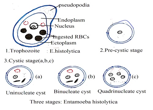
Entamoeba histolytica is a pathogenic protozoa parasite. It exists as Entamoeba histolytica trophozoite, Entamoeba histolytica Precystic stage, and Entamoeba histolytica cystic stage.
1. Morphology of Entamoeba histolytica trophozoite (Growing stage or Feeding stage): Morphology of Entamoeba histolytica trophozoite is as below details.
· Shape: Entamoeba histolytica trophozoite constantly changes its shape.
· Size: 18 to 40 µm
· Cytoplasm: Entamoeba histolytica trophozoite cytoplasm is divided into two portions. These are ectoplasm and endoplasm. Ectoplasm is translucent and endoplasm is granular. Occasionally, endoplasm has red blood cells, leucocytes, and tissue debris.
· Nucleus: Entamoeba histolytica trophozoite nucleus is spherical in shape. The size of the nucleus varies from 4 µm to 6b µm. The nucleus is not present in the center of the Entamoeba histolytica trophozoite cytoplasm. The nucleus is lined with the nuclear membrane.
2. Morphology of Entamoeba histolytica precystic stage (Immature cyst): It is round or slightly ovoid. It has blunt pseudopodium. Red blood cells and other ingested cells are not present in the endoplasm of Entamoeba histolytica precyst. It has a larger nuclear structure than Entamoeba histolytica trophozoite.
3. Morphology of Entamoeba histolytica cyst: In unfavourable condition Entamoeba histolytica trophozoite forms Entamoeba histolytica cyst. It is spherical in shape. Its size varies from 6 to 9 micrometers. It has a clear cytoplasm. Nucleus develops in a cyst in three stages, These are uninucleated cyst (one nucleus), binucleated cyst (two nuclei), and quadrinucleated cyst (Four nuclei) and four nuclei. One cyst forms four trophozoites in favorable conditions.


Entamoeba histolytica Life Cycle
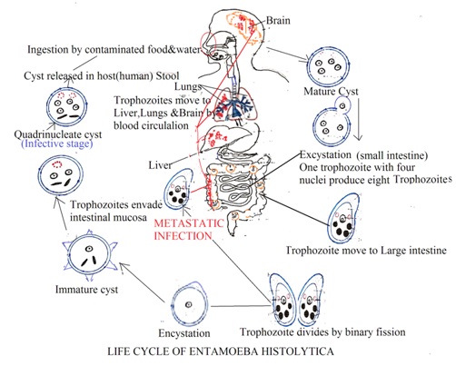
E. histolytic exists as trophozoites, precyst, and cysts. Amoeba cysts can survive inside the human body, outside the human body, and in unfavourable conditions for a longer duration. These cysts are metabolically inactive E. histolytic cells. Trophozoites are metabolically active E. histolytic cells. They are invasive in nature and cannot survive outside the human body.
Amoeba cyst enters the human body through food and water contaminated with amoeba cysts. Faecal matter containing E. histolytic contaminates food and water. Inside the human body, amoeba undergoes through following stages in its life cycle.
1. E. histolytic cysts can also enter the human digestive system through contaminated hands. Cyst becomes active inside the intestine and form trophozoites.
2. These trophozoites multiply inside the intestine by binary fission. Trophozoites invade the mucosa of the large intestine and produce ulceration in the large intestine mucosal wall. Trophozoites from the large intestine may enter into the systemic circulation to produce systemic amoebic infection. Trophozoites infect the liver, lungs, and brain. It is called invasive infection (Metastatic infection).
3. Trophozoites slowly enter the rectum. Inside the rectum, these trophozoites undergo encystations, immature cyst bi-nucleated cysts, and form quadrinucleated cysts. These cysts are excreted out from the body with faecal matter. These cysts contaminate water, food and soil. These cysts enter the human digestive system with contaminated food and water.


Laboratory diagnosis of Entamoeba histolytica
The laboratory diagnosis of protozoa Entamoeba histolytica causes amoebiasis in humans. There are several methods to detect and identify Entamoeba histolytica in the stool sample.
1. Macroscopic examination: Stool color: dark brown, pH: acidic, consistency: viscous fluid with blood and mucus. It is most suitable for acute amebiasis caused by E. histolytica.
2. Microscopic examination: It is the most common and reliable method to detect and identify worms. It is most suitable for
a. Direct wet method: Mix fresh stool with saline solution and examine it under a microscope. Entamoeba histolytica trophozoite and cyst can be visualized under a microscope. Entamoeba histolytica trophozoites have a single irregular nucleus with engulfed RBCs.
b. Concentration technique: Sedimentation technique, floating technique, or formalin ether makes it easy to detect E. histolytica in concentrated stool samples. This technique separates E. histolytica from stool debris.
3. Stool antigen detection:
Enzyme-linked Immunosorbent assays (ELISA): It detects specific antigens of E. histolytica in the stool sample. t is a very sensitive test, ELISA kit is commercially available in the market.
Immunochromatographic test: It is also a sensitive test to detect E. histolytica. It detects specific antibodies of E. histolytica antigen in the stool sample.
4. Molecular technique:
Polymerase chain reaction (PCR): It is a highly sensitive test because it amplifies the DNA of E. histolytica. It can detect even low concentrations of E. histolytica in the stool sample.
5. Serological tests: It is an indirect haemagglutination assay (IHA). This detects the antibodies produced by the E. histolytica. It can detect acute and chronic infections. It is most suitable to diagnose E. histolytica in the liver (Hepatic amoebiasis).
Dr Pramila Singh
