Compound Microscope Handling
Handling of a Compound Microscope. Care and maintenance of different parts of a Compound Microscope. Principle of Working of a Fluorescent Microscope.
MICROBIOLOGY
11/1/20236 min read
Handling of a compound microscope. Care and maintenance of different parts of a compound microscope. Principle of working of fluorescent microscope. Unit IV
Compound microscope
A microscope is a scientific optical instrument that magnifies invisible objects to the naked human eye for their detailed study. There are various types of microscopes like light, compound, electron, etc. A compound microscope is a type of optical microscope that uses multiple lenses to magnify invisible objects. It is called a compound microscope because it uses a combination of lenses to magnify object images.
Definitions
Compound Microscope: A microscope that uses two sets of lenses to magnify small objects is called a compound microscope.
Fluorescent Microscope: A microscope that uses fluorescence to study the properties of organic or inorganic substances is called a fluorescence microscope.
Microscopy: To view objects that cannot be seen with the naked eye by using a microscope is called microscopy.
Magnification: Magnification is the process of enlarging the size of an object. It is carried out by using magnifying glasses, microscopes, or telescopes. The magnification is typically expressed as 2x, 5x, or 10x. 2X means the image appears two times larger than the actual object size. 5X means the image appears five times larger than the actual object size. 10X means the image appears ten times larger than the actual object size.
Resolving Power: Resolving power is the ability of the microscope to distinguish between two closely spaced objects. It is the minimum distance between two points that can be resolved as separate points using a microscope. A higher resolving power means smaller and finer details of the object are visible in the microscope.
Working Distance: The distance between the front lens of the objective and the surface of the specimen when the specimen is in sharp focus is called Working distance. OR The space between the microscope and the object being viewed is called Working distance. This distance depends on the type of objective lens used in the microscope.
Refractive Index: The refractive index indicates how much light bends as it passes from one medium (like air) into another medium (like the lens or the specimen). It determines the ability of a microscope to focus light and create clear images.
Components (Parts) of Compound Microscope:
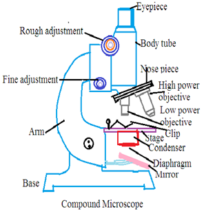
A compound microscope consists of main two types of parts: Mechanical Parts and Optical Parts.
Mechanical Parts:
1. Base: It provides support to make the compound microscope stable on the working top. Its shape is like horse-shoes.
2. Pillar: It is the vertical part to connects the arm and base.
3. Arm (Limb): It is a curve-shaped structure to handle a compound microscope. It supports the stage and body tube of the compound microscope.
4. Inclination joints: It connects the arm and pillar. It is a joint to help adjust the compound microscope for a suitable position.
5. Stage: It is a rectangular platform to hold slides. A hole is present in the center of the platform. Light falls on an invisible object through this hole. The stage has two clips to hold slides and fix the position of the slide on the stage.
6. Body tube: The upper part of the compound microscope arm is attached with a hollow tubular structure. Light passes through the object and enters a hollow tube. Its upper end has a draw tube to hold the ocular lens. Its lower end has a revolving nose piece to hold objective lenses. The body tube is moved up and down by Coarse Adjustment Knob for a longer distance. It helps to focus invisible objects on the slide fixed on the stage of the compound microscope. The body tube has also Fine Adjustment Knob. It moves the body tube up and down for a shorter distance for fine focusing the invisible object.
Optical Parts
7. Reflector: It is a mirror present just above the base of the compound microscope. It reflects light into the condenser, diaphragm, and invisible object on the stage. The reflector has two types of mirrors: plane mirror and concave mirror. A plane mirror is used for bright light sources to reflect and a concave mirror is used for weak light sources to reflect.
8. Condenser: It is present just above the reflector to receive light from the reflector. It can be moved up and down to focus light.
9. Diaphragm: It is present in between the condenser and stage of the compound microscope. It controls the amount of light to passes through an object on the stage of a compound microscope. Diaphragm is of two types: disc diaphragm and iris diaphragm.
10. Objective Lenses: The revolving Nose piece of the compound microscope has three types of lenses Low Power Objective (10X), High Power Objective (45X), and Oil Immersion Objective.
11. Ocular Lens: The draw tube of the body tube has an ocular lens also called the Eyepiece lens. The power of ocular lenses to magnify image are 5X, 10X, 15X, and 20X. The compound microscope that uses one eyepiece at a time is called Monocular Compound Microscope. The compound microscope that uses two eyepieces at a time is called Binocular Compound Microscope.


Handling of the compound microscope:
Handling of the compound microscope requires care and attention to accurate and safe usage of the microscope.
1. Set a compound microscope on a flat, stable surface. Clean the stage of the compound microscope.
2. Decide a power source. It may be natural sunlight or electric light. Adjust light intensity as per requirement.
3. Place the glass slide containing the object on the stage of the compound microscope.
4. Gently turn the body tube of the compound microscope using the coarse adjustment knob to focus the object on the slide.
5. Carefully adjust the slide position to bring the object under an ocular lens.
6. Use a fine adjustment knob for fine and clear focusing of the object on the slide.
7. Adjust light brightness to make the object image clearly visible.
8. If required, select a higher-power ocular lens.
9. Carefully observe the object image.
10. Switch off the electric source after observation, clean the microscope, and place it in the microscope box.
Care and Maintenance of Different parts of Compound Microscope
Proper care and handling of the compound microscope is essential for its performance and longevity. The following steps are followed to care for maintain different parts of the compound microscope
1. Objectives and Ocular Lenses: Always keep lenses dry, clean, and free from debris. Avoid touching for lenses with your fingers. Skin oil damages the lenses and reduces the image quality. Avoid knocking objectives against hard surfaces. Do not allow touching of ocular lenses of the object during up and down movement of the body tube. Always use soft, lint-free cloth or lens paper to clean lenses to avoid scratching lenses. Store lenses in a dust-free environment to prevent dust deposition on lenses.
1. Mechanical Components: Always use soft, lint-free cloth or lens paper to clean, the exterior of the microscope. Be gentle to avoid scratching lenses. Handling: Handle the compound microscope gently. Avoid excessive force on the microscope during handling especially adjustment knobs and stage control knobs. Always move parts of the compound microscope slowly and smoothly. Storage: Keep the compound microscope under dry conditions and store it in a dry place away from direct sunlight and extreme temperatures. Avoid microscope exposure to moisture. Always use a dry cloth to clean the compound microscope. Cover: Properly cover compound microscope using a dust cover, if it is not in use. Preferably place it in the compound microscope box.
2. Illumination System: Turn off the light source, if the compound microscope is not in use. Ensure all electric sources are secure and the cord is not damaged.
Principle of Working of Fluorescent Microscope.
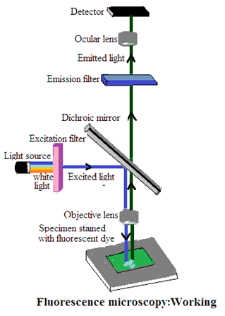
Absorption of shortwave length light like blue light and emission of long wave length light like green and red light is fluorescence. Fluorescent light is used in specialized optical microscope and specialized tools to study microorganisms, biological processes at the cellular and molecular levels, etc. It is widely used in cell biology, microbiology, and pathology. The principle of working of the fluorescent microscope
1. Excitation Light Source: Use a High-intensity lamp to emit a specific short wavelength of light. Normally Ultra Violet lamp is used for the purpose.
2. Filter setting: A fluorescent microscope has a filter in between the light source and the object. The filter allows passing light of the desired wavelength.
3. Specimen Preparation: Fluorophores or fluorescent dyes are used in the sample. They absorb energy from the light of short wavelengths and emit light of longer wavelengths. This produces fluorescence.
4. Emission filter: The second set of filters in a fluorescence microscope is presently called the Emission filter or barrier filter. It selects the wavelength range of emitted fluorescence to pass to the detector. This allows the microscope to capture only the fluorescence emitted by the fluorophores.
5. Detection and visualization: A detector is a charged-couple-device camera or photomultiplier tube. It records the intensity of fluorescence in the sample. It creates a fluorescent image that can be visualized on a monitor for further analysis.
6. Image Processing: Fluorescent microscope has specialized software to enhance contrast and adjust color.
Dr Pramila Singh


