Cardiac Cycle
Cardiac Cycle: Atrial systole – Phase I, Ventricular systole – Phase II and Joint diastole – Phase III. Heart sound, Systolic sound, or first heart sound, Diastolic sound or second heart sound. Cardiac output
Dr Pramila Singh
3/17/20244 min read
HSBTE DMLT HAP IInd Semester Cardiac Cycle: Cardiac Cycle: Atrial systole– Phase I, Ventricular systole – Phase II and Joint diastole – Phase III. Heart sound, Systolic sound, or first heart sound, Diastolic sound, or second heart sound. Cardiac output.
Cardiac Cycle:
The working of the heart can be studied under the cardiac cycle which includes the flow of blood, heartbeat, heart sound, pulse, cardiac output, cardiac impulse ECG, etc.
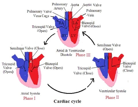
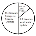
Contraction of the heart chamber (systole) and relaxation of the heart chamber (diastole) constitutes a cardiac cycle. One cardiac cycle stands for 0.8 seconds. One cycle is also known as a heartbeat. During the cardiac cycle, the heart undergoes different phases: Atrial systole – Phase I, Ventricular systole – Phase II, and Joint diastole – Phase III.
1. Atrial systole – Phase I: Blood from different parts of the body enters both atria through their atrioventricular valve. 70% of blood enters into ventricles from the atria without contraction of the atria. The inferior vena cava, superior vena cava, and coronary sinus pour deoxygenated blood into the right atria. The pulmonary veins pour oxygenated blood into the left atria. This stimulates the SA node. Impulse spread from the SA node to both atria walls. Both atria contract (atrial systole) to pour blood into the ventricle (right atria blood into the right ventricle, left atria blood into the left ventricle). Atrial systole lasts for 0.1 seconds.
2. Ventricular systole – Phase II: After completion of atrial systole, the atria dilate for 0.7 seconds. 70% of ventricles filled with blood from atria without effort. The remaining 30% of the ventricle is filled with blood from the atria during atrial systole. SA node impulse reaches AV node at end of atrial systole. AV node transmits the impulse to the ventricle through a bundle of HIS and Purkinje fibers. This initiates ventricular contraction (ventricular systole). Left ventricle contraction pumps blood into the aorta and right ventricle contraction pumps blood into the pulmonary aorta. Ventricular systole lasts from 0.3 seconds. Ventricular contraction opens a semilunar valve in both ventricles and closes the bicuspid and tricuspid valve (tricuspid valve at the right atria ventricular aperture and a bicuspid valve at the left atria ventricular aperture.). This closing produces a heartbeat sound called Lubb
3. Joint diastole – Phase III: Ventricle starts to relax after completion of ventricle systole. It is called ventricular diastole ventricle diastole and lasts for 0.4 seconds. At this period both atria and ventricle relax. There will be no contraction anywhere in the heart. This causes forceful closure of the pulmonary valve and aortic valve. This closure produces dupp sound. Dupp sound is produced at beginning of ventricular diastole.


Heart sound:
Heartbeat produced characteristic sounds. In a normal person, there are two types of heart sounds.
Systolic sound or first heart sound is known as lubb. This sound is due to the closure of the bicuspid valve or tricuspid valve at the beginning of ventricle contractions. It lasts for 0.16 sec with a low-pitched and not very loud sound. It is considered a longer-duration sound.
The diastolic sound or second heart sound is known as dupp. It lasts for 0.10 sec with high pitch and low and sharp sound. It is considered as a short-duration sound. It is due to the closure of the pulmonary valve and aortic valve at the beginning of ventricular systole. The heart sound is also called the Lubb-Dupp sound.
Sound quality helps to predict the health of the heart. In case of valve injury, this sound will be lubb shh. It is known as a heart murmur.
Cardiac output: The amount of blood pumped by the heart in one minute is called the cardiac output. A normal heart pumps 70 ml of blood on each ventricular contraction. The average heartbeat is 72 per minute. It means the heart pumps 72 X 70 ml of blood per minute which is 5.04 L. Cardiac output is almost equal to the total volume of blood present inside the body.
Nerve supply to the heart:
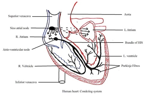
The heart has two nodes; these are the sinoatrial nodes (SA Nodes) and the atrioventricular node (AV node). It lies in the right atrium just below the superior vena cava opening. Fibers arise from the SA Node and encircle the right atrium and left atrium. Through fibers, the electric impulse passes to the right atrium and left atrium wall.
The AV node is present in the right atrium septum wall near the coronary sinus opening. It stimulates electric impulses received from the SA node. A Bundle of HIS arises from the AV node passes through the atrioventricular septum and enters into the interventricular septum. The bundle of HIS forms the right and left branches. Each branch produces various small branches. These branches are also called Purkinje fibre. These nerve fibers carry impulses to contact heart muscles.


