Blood Glucose Estimation
HSBTE DMLT IInd Semester Blood glucose estimation, screening test and glucose tolerance test (GTT), Principle and methods of estimation, Reference values, Renal threshold, Clinical importance of blood sugars/GTT
BIOCHEMISTRY
Dr Pramila Singh
3/20/202410 min read
HSBTE DMLT IInd Semester Blood glucose estimation, Screening Test and Glucose Tolerance Test (GTT), Principle and Methods of Estimation, Reference Values, Renal Threshold, Clinical Importance of blood sugars/GTT.
Blood glucose estimation, screening test, and glucose tolerance test (GTT/OGTT).
PRINCIPLE and methods of glucose estimation:
Protein-free filtrate of whole blood is used as a specimen to estimate blood glucose. This specimen gives a lower value of glucose than the plasma specimen. It is mainly due to the presence of RBCs in whole blood. RBC metabolic activity decreases the concentration of glucose in the protein-free filtrate of whole blood. Most of the methods recommend plasma as a specimen, not serum or whole blood. Plasma or whole blood as a specimen requires an anticoagulant in the specimen. Fluoride-oxalate mixture (2mg sodium fluoride and 6 mg potassium oxalate per ml of blood) is the most preferred anticoagulant in specimens to estimate blood glucose. Fluoride acts as an anticoagulant and also inhibits glycolysis of glucose present in the specimen. Chemical Formula for Glucose is C6H12O6. Molecular weight of glucose is 180.16 grams per mole (g/mol).
Glucose in blood plasma and serum is determined by the following methods: Ortho-toluidine method, Glucose oxidase method, Folin-Wu method, and Reflometric method.
GOD POD ( Glucose oxidase Peroxidase method) is the best laboratory method to estimate blood glucose.
Advantages of GOD-POD Method:
High Specificity: GOD only reacts with glucose, minimizing interference from other substances in the blood, unlike o-toluidine and Folin-Wu methods.
Accuracy: The method offers excellent accuracy in measuring blood glucose levels.
Sensitivity: It can detect even small changes in glucose concentration.
Linearity: The relationship between color intensity and glucose concentration is linear within a specific range, simplifying calculations.
Disadvantages of o-Toluidine and Folin-Wu Methods:
Non-Specificity: These methods react with various reducing sugars in the blood, not just glucose, leading to inaccurate results.
Lower Accuracy: Measurements can be less precise compared to the GOD-POD method.
Other Limitations: They may require additional steps or have limitations in sensitivity and linearity.
1. Ortho-toluidine method (O-toluidine method).
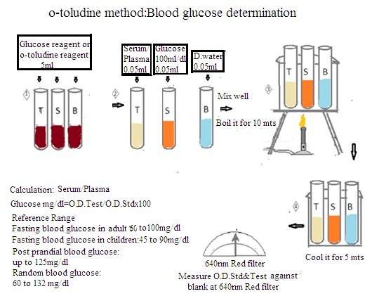
Principle: It is a colorimetric method. Glucose reacts with Ortho-toluidine in the presence of glacial acetic acid at 100 degrees C to produce a green color complex. The intensity of color is directly proportional to the concentration of glucose in the blood specimen. Color intensity is measured using a photometer at 620 nm to 660 nm.
This method does not require the deproteinization of blood specimens to perform tests. However, this method is not used due to the carcinogenic properties of Ortho-toluidine.
Procedure:
i. Blood specimen collection: Three types of blood samples can be collected. These are:
a. Fasting specimen (P): Collect 2 to 3 ml blood after 12 to 16 hrs fasting in a fluoride bulb.
b. Post glucose specimen (PG): Administer orally 5gm glucose after Collection of fasting blood specimen. Collect 2 to 3 ml blood specimen after 2 hours.
c. Post parandial specimen (PP): 2 to 3 ml blood specimen is collected in a fluoride bulb after 2 hours of lunch.
d. Blood serum is used for the test. The serum should be separated from clotted blood within 30 minutes after blood collection.
ii. Reagents preparation: Ortho-toluidine reagent is prepared by mixing 60 ml Ortho-toluidine, 940 ml glacial acetic acid, and 1.5 gm thiourea. It is stored in an amber color bottle at 2-8○C. It remains stable for up to 6 months.
iii. Glucose standard: Dissolve 1 gm glucose in 100 ml saturated solution of benzoic acid (0.3%).
Procedure:
Label three test tubes (100mm x10mm) as test, standard, and blank. Dispense reagents and serum as per the below table detail
Mix them properly and boil in a water bath for 10 minutes. Allow to maintain room temperature. and Measure the optical density of the standard and test against blank.
Calculations: Glucose, mg/dl = O.D.Test/O.D.Std X 100.


2. Glucose-oxidase method (Oxidase-peroxidase method or God and Pod Method)
God and Pod Method is the best laboratory method to estimate blood glucose level
principle: It is the enzymatic determination of glucose in blood serum. Glucose Oxidase is an enzyme.
· Glucose oxidase oxidizes the aldehyde group of glucose to produce gluconic acid and hydrogen peroxide.
· Enzyme peroxidase breaks hydrogen peroxide into water and oxygen.
· The released oxygen reacts with 4-aminophenazone and phenol to produce the pink-coloured compound.
· The intensity of color is measured by using a photometer at 530 nm.
Reagents:
Protein precipitant: Make a solution of sodium tungstate 10gm, disodium hydrogen phosphate 10gm, and sodium chloride 9gm in 400ml distilled water. Adjust PH 3.0 by adding 125 ml of 1.0 ml HCl.Use phenol 1gm as a preservative. Add sufficient distilled water to produce a volume of 1 liter.Storeat4o C.
i. Phosphate buffer, pH 7.0: Make a solution of disodium hydrogen phosphate 8.52gm and potassium dihydrogen phosphate 5.44gm in 500ml distilled water. Adjust pH 7.0 and add sufficient distilled water to produce a volume of 1 liter.
ii. Buffer enzyme (Colour reagent): Prepare color reagent by using the following formulation:
a. Glucose oxidase: 650 units
b. Peroxidase: 500 units
c. 4-aminophenazole: 20mg.
d. Sodium azide: 30mg.
e. Phenol: 0.1 gm
f. Distilled water to produce a volume of 100ml
iii. Glucose standard, solution: 1gm glucose, 0.3% benzoic acid in 1000ml distilled water. (100mg/dl).
Procedure: Procedure:
Label three test tubes (100mm X 10mm) as test, standard, and blank. Dispense reagents and serum as per the below table details.
Mix them properly and set aside at room temperature (37 degrees C) for 30 minutes. Measure the optical density of the standard and test against blank at 530 nm (red filter).
Calculation: O.D Test/ O.D. Standard x 100.

Important terms related to Blood Glucose
1. Glycolysis: Metabolism of glucose in the cell's cytoplasm to form pyruvate and lactate with the generation of ATP (Adenosine tri phosphate) and NADPH.
2. Glycogenesis: The conversion of glucose to glycogen for storage in the liver and muscles is called glycogenesis.
3. Glycogenolysis: The breakdown of glycogen into glucose is called glycogenolysis. It occurs mainly in the liver and muscles. It releases glucose into the blood to maintain blood glucose levels.
4. Glyconeogenesis: The synthesis of glucose inside the human body from a non-carbohydrate source is called glyconeogenesis. It maintains blood glucose levels during fasting or low carbohydrate intake.
3. Folin-Wu method:
It is also known as the reduction of the cupric method to the cuprous salt method. It is the oldest method and is not in use.
Principle:
It uses the following three reactions
i. Blood protein precipitation by using copper tungstate.
ii. Cupric sulfate reduction to cuprous oxide
iii. Combination of cuprous oxide with molybdate reagent to develop blue-green color.
Reagents:
a. Sodium tungstate 10% solution in distilled water
b. 2/3 N Sulphuric acid
c. Alkaline copper tartrate reagent:
a. Solutions of sodium carbonate 40gm, tartric acid 7.5gm in 400ml distilled water.
b. Solution of 4.5 gm copper sulfate in 100 ml of distilled water.
c. Mix both solutions and add sufficient water to make a volume of 1000 ml.
d. Phosphomolybdic acid reagent: Solutions of sodium tungstate 5gm, molybydic acid 35gm, 10% sodium hydroxide 200ml in 200ml distilled water. Boil the solution for 45 minutes to remove ammonia. Cool it and add 89% phosphoric acid 125 ml. Add sufficient distilled water to make volume up to 500ml.
e. Glucose standard solution 100mg/dl with benzoic acid.
Procedure:
Use protein-free whole blood filtrate to measure optical density.
Calculation: O.D Test/ O.D. Standard x100.
Reducing sugars and non-reducing sugars are two categories of carbohydrates.
Reducing Sugars:
Definition: A reducing sugar is a carbohydrate that can donate electrons to another chemical species in a redox chemical reaction.
Reactivity: It can reduce other substances, such as copper ions (e.g., in Benedict's or Fehling's solution), resulting in the formation of a colored precipitate.
Examples: Glucose, fructose, lactose, and maltose are examples of reducing sugars.
Biological Significance: Many reducing sugars play essential roles in biological processes such as energy production and storage.
Non-Reducing Sugars:
Definition: A non-reducing sugar is a carbohydrate that cannot donate electrons and thus cannot reduce other substances.
Reactivity: It does not react with oxidizing agents like Benedict's or Fehling's solution.
Examples: Sucrose is the most common example of a non-reducing sugar.
Biological Significance: Non-reducing sugars are important energy sources and structural components in organisms.


4. Reflometric method Principle:
A reflocheck meter is used to measure glucose in a given sample. A reagent strip is used that contains oxidase peroxidase enzymes and color reagent.
Procedure:
Put capillary blood on the reagent strip. Insert it in the reflocheck meter. It will give a reading of glucose in the blood in mg/dl.
Reference value:
1. Fasting blood glucose level for adults: 60 to 100mg/dl
2. Fasting blood glucose level for children: 45 to 90mg/dl
3. Postprandial blood glucose: up to 125mg/dl
4. Random blood glucose: 60 to 132 mg/dl.
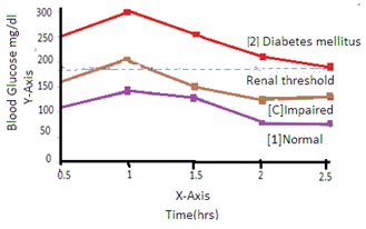
Renal threshold: Renal glucose threshold is the blood plasma glucose level at which glucose appears in urine. The presence of glucose in the urine is termed glycosuria. The renal threshold value is variable. It depends upon the physiological condition of the patient. It helps to predict diabetes in patients.
Screening test:
A screening test to tolerate glucose inside the body is performed before the glucose tolerance test. The following points are considered in the screening test.
1. Glycosuria: The appearance of glucose in urine is called glycosuria.
2. Diabetes: without glycosuria.
3. Genetics: family history of diabetes.
4. Neuropathy and retinopathy: neuropathies and retinopathies of unknown reason.
5. Pregnancy, liver disease, infection or thyrotoxicosis:
There are two main types of screening tests for blood glucose, depending on whether you need to fast beforehand:
1. Fasting Blood Sugar (FBS) Test:
This is the most common screening test for diabetes and prediabetes.
It requires fasting for 8-12 hours before the blood draw.
During fasting, the body relies on stored glucose for energy. So, an elevated blood sugar level after fasting indicates problems with glucose regulation.
2. Random Blood Glucose (RBG) Test:
· This test doesn't require fasting.
· It can be used as a quick initial screening tool, but a high RBG result needs to be confirmed with an FBS test or other follow-up tests for a definitive diagnosis.
Interpretation of Results:
· Normal FBS level: Generally below 100 mg/dL (5.6 mmol/L).
· Prediabetes: FBS level between 100-125 mg/dL (5.6 - 6.9 mmol/L).
· Diabetes: FBS level of 126 mg/dL (7.0 mmol/L) or higher on two separate tests.
Glucose Tolerance Test (GTT):
The glucose tolerance test is also known as the oral glucose tolerance test (OGTT). The ability of the body to utilize glucose present in blood circulation is called glucose tolerance. Glucose tolerance depends upon the rate of glucose absorption and the release of insulin into blood circulation. A decrease in the release of insulin into blood circulation decreases glucose tolerance in the body. The following steps are followed in GTT:
1. Patient preparation:
· Instruct patient to consume at least 150gm carbohydrate for three continuous days before GTT date.
· Instruct patient to not eat or drink anything except water on GTT date.
· Instruct patient not to exercise or long walk or fear on GTT date.
· Perform GTT in the morning hour.
Procedure:
· Administer 75 gm or 100gm glucose orally with 300 ml of drinking water.
· Note down the time
· Collect four blood samples at an interval of 30-minute intervals for 2 hours after glucose administration
· After 2 hours collect at least two urine samples at intervals of 60 minutes.
Result:
Determine blood glucose and urine glucose levels. Prepare a graph by putting time on the X-axis and blood plasma glucose on the Y-axis. The curve plotted on the graph is called the glucose tolerance curve.
Clinical importance of blood sugar/GTT:
The glucose tolerance test (GTT) is used to assess glucose (sugar) metabolism in the human body. It is used in the diagnosis and management of diabetes mellitus.
The following are the clinical importance of the glucose tolerance test:
1. Diagnosis of Diabetes Mellitus: GTT is a diagnosis of diabetes mellitus. It helps in identifying individuals with impaired glucose tolerance or impaired fasting glucose. These are pre-diabetic states. Early detection allows for timely treatment management to prevent the progress of diabetes and its complications.
2. Assessment of Insulin Resistance: Insulin resistance is a condition where the body's cells become resistant to the effects of insulin. It leads to elevated blood glucose levels. GTT helps in assessing insulin sensitivity and identifying individuals at risk of developing type-2 diabetes.
3. Monitoring Gestational Diabetes: Some women develop gestational diabetes during pregnancy. GTT is used for screening and diagnosing gestational diabetes. It is important for the health of both the mother and the baby. Early detection and management of gestational diabetes reduce the risk of complications during pregnancy and childbirth.
4. Evaluation of Hypoglycemia: GTT is used to evaluate hypoglycemia (low blood sugar levels). Healthcare providers can assess the body's response to glucose intake and identify abnormalities in glucose metabolism that may contribute to hypoglycemic episodes.
5. Assessment of Beta Cell Function: Beta cells in the pancreas are responsible for producing insulin. GTT provides valuable information about beta cell function by assessing the pancreas' response to a glucose challenge. This information is important to understanding the pathophysiology of diabetes and guiding treatment decisions.
6. Risk Stratification for Cardiovascular Disease: Individuals with impaired glucose tolerance or diabetes have an increased risk of cardiovascular disease. GTT can help identify individuals at higher risk, allowing for early lifestyle modifications to reduce the risk of cardiovascular complications.
Symptoms of Diabetes
Frequent Urination (Polyuria): Increased urination to eliminate excess glucose through urine.
Excessive Thirst (Polydipsia): Frequent urination leads to thirst.
Unexplained Weight Loss.
Fatigue: Fatigue and weakness occur.
Blurred Vision: High blood sugar levels lead to blurred vision.
Slow Healing of Wounds
Frequent Infections: More prone to infections.
Increased Risk of Gum Disease.
Dr Pramila Singh


Factors affecting Blood Glucose Level (Regulation of Blood Glucose)
Blood sugar regulates energy to cells. The pancreatic islets in the pancreas secrete Insulin and Glucagon to regulate blood glucose levels.
· Insulin: It is a "blood sugar regulator". It is produced by beta cells in the pancreas. The pancreas releases insulin when blood sugar rises. Insulin allows glucose to enter into cells.
· Glucagon: It is produced by alpha cells in the pancreas, It acts against insulin. The pancreas releases glucagon when blood sugar levels drop too low. Glucagon allows releasing glucose into the bloodstream. It raises blood sugar levels.
· Liver: The liver acts as a storage unit for excess glucose as glycogen. Liver breaks glycogen into glucose (Glycogenolysis) when blood sugar levels are low. It maintains blood sugar balance.
· Kidneys: The kidneys remove excess glucose from the bloodstream through urination.
· The Digestive System: Simple carbohydrates are broken down quickly. It causes blood sugar spikes. Complex carbohydrates are absorbed more slowly. It leads to more gradual increases in blood glucose levels.
· Hormones: Several other hormones, such as cortisol (stress hormone) and epinephrine (adrenaline) also influence blood sugar levels. They increase glucose production or decrease insulin sensitivity.
