Automation in hematology
Automation in hematology: Various types of Blood cell counter. Principle and operation of the automated blood cell counters. IInd Semester DMLT Unit V.
Dr Pramila Singh
5/1/20245 min read
Automation in hematology: Various types of Blood cell counter. Principle and operation of the automated blood cell counters. IInd Semester DMLT HSBTE Unit-V.
AUTOMATED BLOOD CELL COUNTERS (HAEMATOLOGY CELL COUNTER)
Automation in hematology counts blood cells more accurately. For this purpose, a blood cell counter (hematological cell counter) is used to measure hematological parameters: white blood cell count, hemoglobin, red cell indices, etc. Blood cell counters are used for the following purposes
Identification of blood cells: Red Blood cells (Erythrocytes). White Blood Cells (Leucocytes) and platelets.
Identification of subclasses of blood cells such as granulocytes, and agranulocytes. lymphocytes etc.
Determination of blood cell numbers and blood cell size.
Types of blood cell counters: Fully automated and semi-automated blood cell counters are available. Their selection depends upon the parameters of the blood to be measured. There are mainly two types of automated blood cell counters
A. Coulter Blood Cells counter
B. Sysmex Blood cells counter
C. Flow cytometry blood cell counter
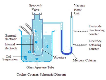
COULTER BLOOD CELLS COUNTER (Coulter analyzer): It consists of a glass tube. It has a small orifice on its wall near its closed end. The diameter of the orifice is 100 micrometers. This glass tube is called a glass aperture tube. It holds a diluting fluid that is a good conductor of electricity such as normal saline water. Normal saline water contains 0.85 gm of sodium chloride in water as a solution. This means Normal saline water is 0.85% sodium chloride solution in water. The Glass tube also holds an electrode dipped in fluid. This electrode is called an internal electrode.
The lower half part of the glass aperture tube is immersed in a container. This container holds blood cells suspended in a diluent fluid such as normal saline water. The container also holds one electrode. That is called an external electrode. The wall of the glass aperture tube acts as an insulator between the internal electrode and the external electrode.
The open end of the glass aperture tube has a stopcock valve to close the glass aperture tube. There is one aperture on the wall of the glass tube near the stop cock valve. This aperture is connected with a “U” shaped glass tube. “U” shaped glass tube glass tube is partly filled with mercury. There are two electrical points on the right limb of the “U” shaped glass tube. These electric points are attached to electrodes Upper electric point acts as the electrode deactivating counter and the lower electric point acts electrode activating counter. The open end of the “U” shaped glass tube right limb is connected to a vacuum pump.


Principle: Coulter blood cell counter utilizes electrical impedance (resistance) to count blood cells. Electrical impedance is opposition to electric flow. The principle of the Coulter blood cell counter is also called the Coulter principle
Blood cells are poor conductors of electricity. Diluents containing electrolytes are good conductors of electricity. Blood cells present inside diluents shall decrease electric current flow through diluents. Change in electric current flow is measured as a voltage pulse. Voltage pulse is proportional to the number of blood cells present in the diluents. The magnitude of the voltage pulse is proportional to the size of blood cells.
Colter blood cells counter-register voltage pulse and magnitude of each pulse that is printed as a histogram.
Diluents to be used in the Coulter blood cells counter are isotonic solutions. The isotonic solution does not cause any change in blood cell morphology. Normal saline solution (0.85% W/V sodium chloride solution in water) is the most commonly used diluent. It is an isotonic solution.
Operation (Working): Apply vacuum on a “U” shaped glass tube by using a vacuum pump. This will pull mercury up in the right limb of the “U” shaped glass tube. This will force blood cells suspended in a container to enter the glass aperture tube through the orifice. Blood cell suspension shall displace an equal volume of normal saline solution in the glass aperture tube. This will decrease the electric current flow through normal saline solution and create a voltage pulse (impedance). A decrease in the electric current flow of normal saline solution shall be proportional to the concentration of blood cells. The number of voltage pulses shall be equal to several blood cells.
The counting of the electric pulse starts after the mercury level in the right limb of the glass aperture tube touches the electrode activating counter. There will be no counting of electric pulse after the mercury level in the right limb of the glass aperture tube touches the electrode deactivating counter.
SYSMEX BLOOD CELLS COUNTER:
It consists of a transducer and a transducer chamber. The transducer holds an electrode called an internal electrode. The Transducer chamber holds an electrode called an external electrode. The transducer is attached to a vacuum pump. Transducer chamber opens into the transducer through a small orifice. The instrument has also a volumetric manometer filled with diluents. It has an internal ball that floats on diluents.
Operation (Working): Deliver diluted sample in transducer chamber. Apply vacuum in the transducer by using a vacuum pump. Blood cells will enter the transducer from the transducer chamber through the orifice. It will interrupt the current flow between the internal and external electrodes. Current flow interruption shall be directly proportional to blood cell size.
The vacuum pulls the sample from the transducer chamber into the transducer at the same time it will also move the internal ball floating in the volumetric manometer. This float acts to stimulate and stop the sensor from counting current flow. This counting creates a pulse that can be recorded as a histogram.
FLOW CYTOMETRY BLOOD CELL COUNTER:
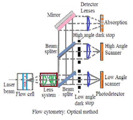
It consists of a laser beam source, a flow cell that holds diluted blood, two types of light sensors, photodetectors, and a computer system to process electrical impulses. One light sensor detects a laser beam that scatters at 180o from the light source. Another light sensor detects a laser beam that scatters at 90o from the light source.
Principle: It is an optical method to count blood cells. Blood cells scatter laser beams The Angle of laser beam scattering depends upon the structure of blood cells. The amount of laser beam scattering at 180o from the laser beam source is proportional to blood cell volume and blood cell density. The amount of laser beam scattering at 90o from the laser beam source is proportional to cellular contents and granules present inside blood cells Photo detectors convert’ scattered laser beams into electric impulses. These electric impulses are processed by a computer.
Fluorochrome dye is used to enhance blood cell identification. It also helps to detect antigens present on the surface of blood cells. Various types of Fluorochrome dye are available. They act on different wavelengths of light. They are useful in differential counts of WBCs.
Operation (Working): Laser beam is passed through flow-cell from laser beam source. A stream of single cells of blood diluted with normal saline solution is placed into a flow cell. Blood cells will scatter the laser beam. Scattering of laser beam shall be detected by photodetectors. Photodetectors convert laser beam signals into electric impulses. Electric impulses shall be processed by a computer to provide blood cell count data.
Care:
It is a sensitive instrument. Install it in controlled humidity, controlled temperature, and free from dust,
Electronic components are sensitive to voltage fluctuation. The electric supply should be through the stabilizer. There should not be sparking in the electric supply plug.
Well-trained and educated staff should be allowed to handle instrument
Stick with manufacturers' instructions/recommendations.
Instruments should be serviced by trained and authorized personnel recommended by the manufacturer.
If the instrument is not in use, unplug its electric supply and keep the instrument under covered conditions.
Applications: Several hematological parameters like white blood cell count, hemoglobin, red cell indices, differential blood count, platelets, etc.
Dr Pramila Singh


