Ascaris lumbricoides
Morphology, Life cycle, and Lab diagnosis of Ascaris lumbricoides
PARASITOLOGY
Dr Pramila Singh
10/15/20235 min read


Morphology of Ascaris lumbricoides (Roundworm or Giant intestinal roundworm)
Ascaris lumbricoides is a parasitic roundworm found in human intestine, pig intestine, and gorilla intestine. Ascaris lumbricoides found in humans and pigs are morphologically similar but biologically dissimilar. They are nematodes belonging to the phylum Nemathelmenthes. Roundworm exists as adult worm and eggs.
Morphology of Adult roundworm (Ascaris lumbricoides)
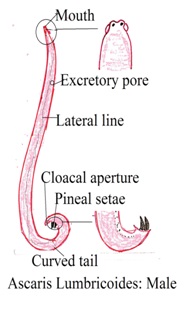
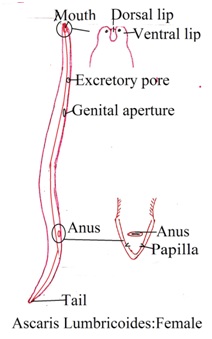
1. Size and shape: It is cylindrical in shape pointed at both ends. The anterior end is thinner than the posterior end. Its color varies from white to pink. Its body has four streaks. The length of a female roundworm varies from 35 cm to 40 cm. The length of a male roundworm varies from 15 cm to 30 cm.
2. Body wall: Its body is covered with a tough and flexible cuticle. The cuticle protects the roundworm from the host immune system and the host digestive system. The cuticle is smooth and resistant to chemical and mechanical stress. Its body has four streaks: two on the lateral side of the body, one on the ventral surface of the body, and one on the dorsal surface of the body.
3. Body cavity: Roundworm has a body cavity filled with body fluid. Body fluid has irritating property that continuously irritates the host intestine.
4. Mouth: The mouth of a roundworm is located at the anterior end of the body. It has three lips that develop a triangular opening of the mouth. Lips help roundworms attach to the host intestine wall and suck semisolid feed from the intestine wall.
5. Digestive system: Roundworm has a complete digestive system consisting of mouth, pharynx, intestine and anus. Posterior part of roundworm after anus
6. Reproductive system: Male roundworm and female roundworm exists separately. The male reproductive system has two testes, vas deferens, a seminal vesicle, and a pair of copulatory parts. The female reproductive system has two ovaries, an oviduct, and one uterus. Eggs develop inside the uterus. Reproductive organs float in the body fluid of roundworms.
7. Excretory system: The excretory system consists of two excretory canals located in the body cavity. One on each side of the roundworm body. The anterior end of the roundworm body has an opening for excretory canals. Roundworm excretes ammonia and urea as excretory products.
8. Nervous system: Roundworm has a simple nervous system. It forms a nervous ring around the pharynx and longitudinal nerve cord along with the length of the body.
9. Respiration: The mode of respiration in roundworms is anaerobic respiration.
Morphology of Egg of Roundworm (Ascaris lumbricoides)
Female roundworm releases both fertilized and unfertilized eggs. Eggs are excreted from the human body with stool. The size of the fertilized eggshell is smaller than the fertilized eggshell. Fertilized eggs are oval while unfertilized eggs are elliptical in shape. Roundworm unfertilized eggs do not float in the saline solution while fertilized eggs float in the saline solution. Fertilized eggshell has a regular coating of albumin while unfertilized eggshell has an irregular coating of albumin.
1. Size and shape: Roundworm eggs look like cylindrical or oval-shaped round at both ends. Their lengths vary from 20 to 100 micrometers.
2. Outer covering: Roundworm eggs have a covering called an eggshell. Eggshell protects the embryo present inside the roundworm egg. The color of eggs depends upon their stage of development. Color may be translucent, yellow, brown, or dark.
3. Egg contents: Eggs contain developing embryo.
Life cycle of Roundworm (Ascaris lumbricoides)
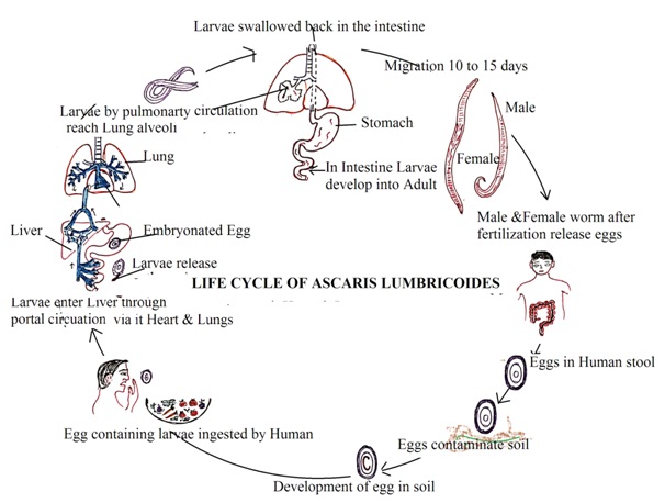
Human is the definitive host (primary host) of Roundworm. However, it requires the human body and a favorable environment outside the human body to complete its life cycle. It has only one host and does not require any intermediate host. The following are the stages of the roundworm life cycle.
1. Stage 1 (Egg stage): Infected human excretes both fertilized and unfertilized roundworm eggs. Unfertilized eggs and fresh fertilized eggs are not infective to humans. Roundworm eggs are resistant to harsh environments.
2. Stage 2 (Larvae stage: Fertilised eggs develop larvae in moist and warm soil. Fertilized eggs have coiled embryos. Larvae development requires 10 to 40 days depending upon humidity and temperature of soil. Larvae go through several stages and shed their outer covering.
3. Stage 3 (Infective stage): At this stage, larvae are capable of developing an infection in the new host. Some species of larvae enter the host body through penetration into the host skin. Some species of fertilized eggs enter into the new host body through the ingestion of contaminated food. Digestive juice in the duodenum digests outer egg shells. Larvae are released from the eggs
4. Stage 4 (Larvae migration): Larvae penetrate the duodenum wall and enter the iver through portal blood circulation. They migrate to the heart, pulmonary circulation, and lungs alveoli through blood circulation. Their size increases in the lung's alveoli. Roundworm larvae enter the respiratory tract from the lung's alveoli and are swallowed into the digestive system with cough and pharynx mucus.
5. Stage 5 (Adult stage): Larvae grow into male and female adult roundworms inside the duodenum. Adult roundworms are sexually active. Male and female roundworms mate to develop eggs inside female roundworms. Female roundworms produce a large number of eggs. Eggs are excreted from the infected host body into a stool. The life cycle of a roundworm inside the human body requires about 2 months.


Laboratory diagnosis of Roundworm (Ascaris lumbricoides)
Roundworm eggs and larvae are detected and identified in the laboratory to diagnose hookworm infection. The number of eggs present in the stool sample decides the intensity of Roundworm infection. The administration of anthelmintic drugs promotes the excretion of worms in stool. Administration of anthelmintic drugs at bedtime and collection of stool samples in the morning is the most suitable technique to detect and identify parasites. This makes the detection easy in stool samples. The following methods are followed to diagnose roundworm infection in the medical laboratory.
1. Stool examination (Egg detection): It is the most common and reliable method to detect and identify worms
Direct wet method: A small amount of fresh stool sample is missed with saline solution and examined under a microscope for the presence of round eggs in the sample.
Concentration techniques such as sedimentation method, floating method, or formalin ether sedimentation techniques are used to concentrate the sample. Concentration techniques increase the chance of detection of roundworm eggs and larvae in the sample.
2. Serological test (Enzyme-linked immunosorbent assays ELISA): ELISA test can detect specific antibodies to roundworms in the blood sample. However, this method is not able to detect acute roundworm infection in humans.
3. Imaging technology: X-rays and ultrasounds are used to visualize adult roundworms in the intestine and other organs. Ultrasonography also reveals the roundworm movement and roundworm clusters in the small intestine.
Dr Pramila Singh
